Copyright
©The Author(s) 2025.
World J Nephrol. Jun 25, 2025; 14(2): 103027
Published online Jun 25, 2025. doi: 10.5527/wjn.v14.i2.103027
Published online Jun 25, 2025. doi: 10.5527/wjn.v14.i2.103027
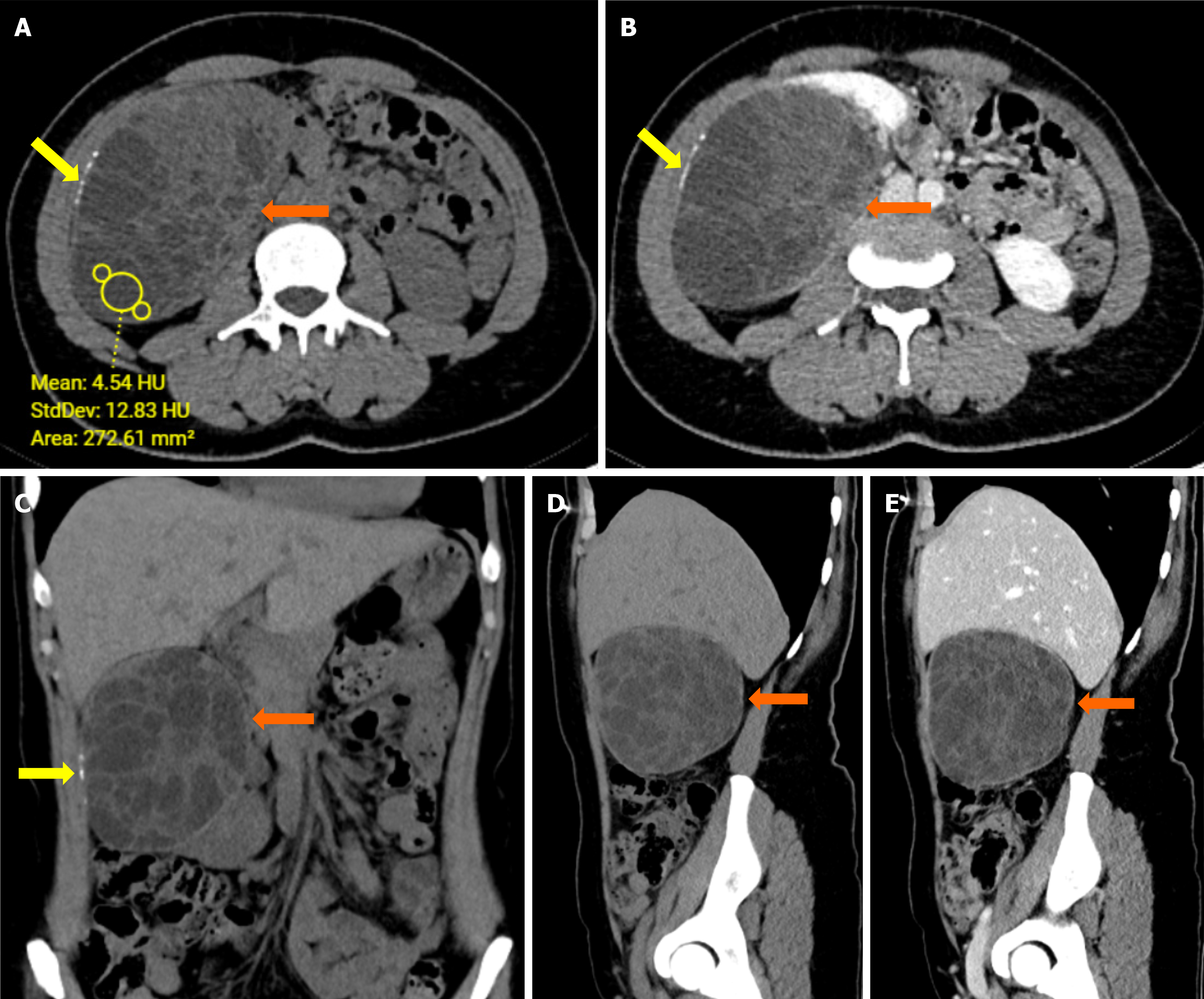
Figure 1 Axial and coronal abdominal computed tomography images of a complex cystic lesion in the right kidney.
A: Axial pre-contrast computed tomography (CT) image shows a complex cystic lesion with several septations in the right kidney (orange arrows); B: Axial post-contrast CT image demonstrates enhancement of the lesion, with noticeable septations (orange arrows) and linear calcifications along its wall (yellow arrows); C: Coronal pre-contrast CT image highlights the extent of the cystic lesion and linear calcifications (yellow arrows); D: Sagittal pre-contrast CT image presents the multiloculated nature of the lesion; E: Sagittal post-contrast CT image further delineates the lesion’s characteristics with contrast enhancement of the septations.
- Citation: Celik AS, Yosunkaya H, Yayilkan Ozyilmaz A, Memis KB, Aydin S. Echinococcus granulosus in atypical localizations: Five case reports. World J Nephrol 2025; 14(2): 103027
- URL: https://www.wjgnet.com/2220-6124/full/v14/i2/103027.htm
- DOI: https://dx.doi.org/10.5527/wjn.v14.i2.103027


