Copyright
©The Author(s) 2025.
World J Nephrol. Jun 25, 2025; 14(2): 100530
Published online Jun 25, 2025. doi: 10.5527/wjn.v14.i2.100530
Published online Jun 25, 2025. doi: 10.5527/wjn.v14.i2.100530
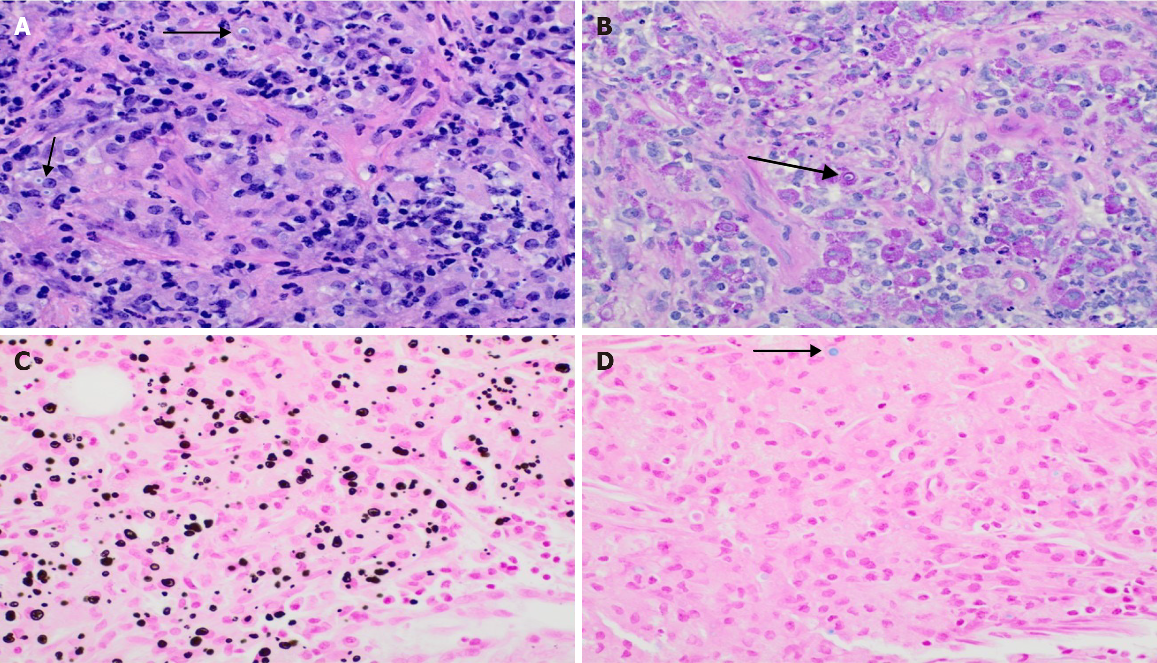
Figure 1 Von Kossa stain.
A: Variable lymphoplasmacytic infiltrate and sheets of macrophages were noted on the haematoxylin and eosin stain(marked with arrows); B: Periodic acid Schiff (PAS) stain highlighting PAS positive intracytoplasmic inclusions (Michaelis-Gutmann bodies) marked with an arrow that focally has a targetoid appearance; C: Von-Kossa stain highlighting the mineralized cytoplasmic inclusions; D: Prussian blue stain for iron that is focally positive(marked with an arrow).
- Citation: Simhadri PK, Contractor R, Chandramohan D, McGee M, Nangia U, Atari M, Bushra S, Kapoor S, Velagapudi RK, Vaitla PK. Malakoplakia in kidney transplant recipients: Three case reports. World J Nephrol 2025; 14(2): 100530
- URL: https://www.wjgnet.com/2220-6124/full/v14/i2/100530.htm
- DOI: https://dx.doi.org/10.5527/wjn.v14.i2.100530


