Copyright
©The Author(s) 2024.
World J Nephrol. Dec 25, 2024; 13(4): 98932
Published online Dec 25, 2024. doi: 10.5527/wjn.v13.i4.98932
Published online Dec 25, 2024. doi: 10.5527/wjn.v13.i4.98932
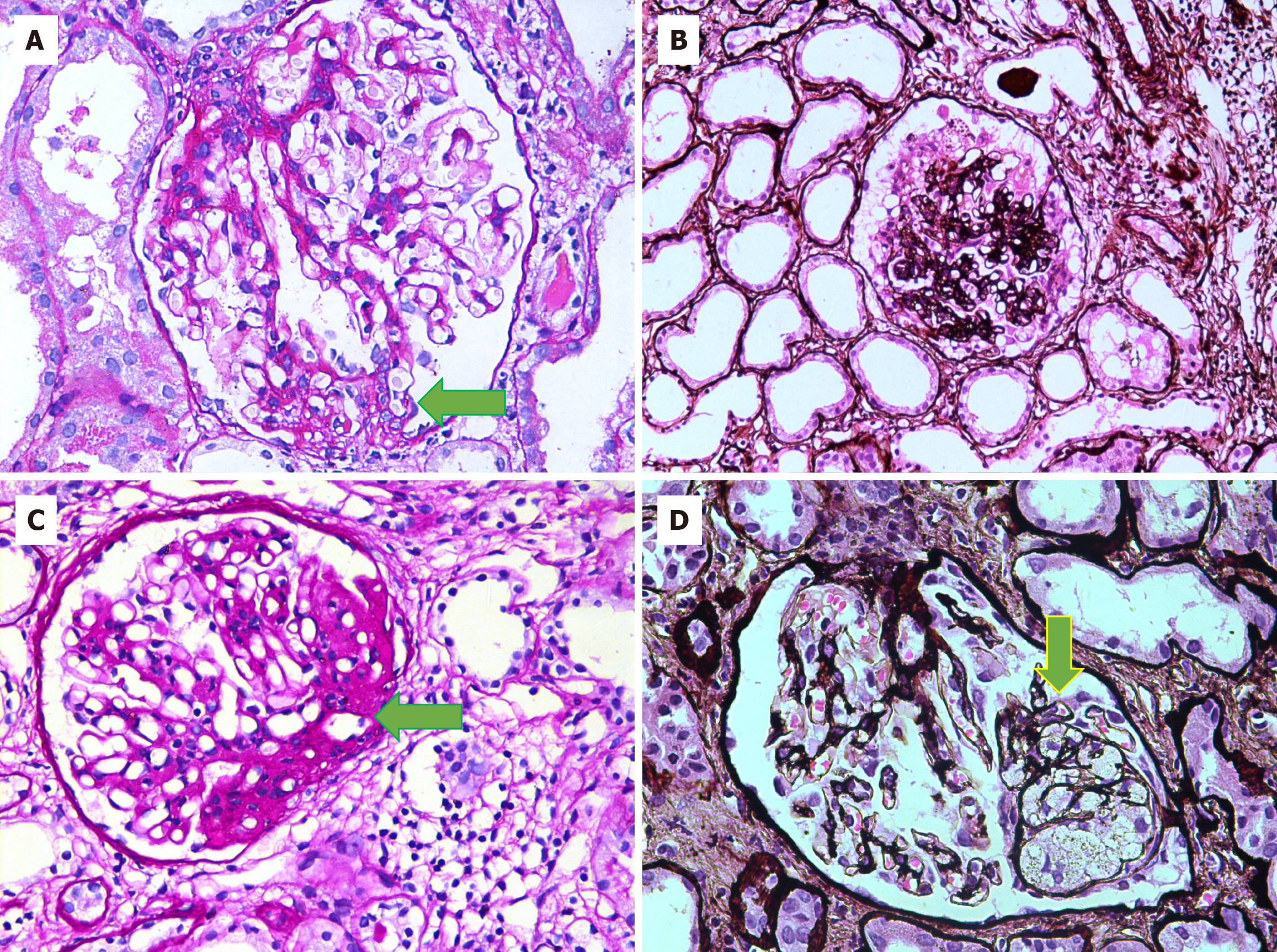
Figure 2 Histopathological features of focal segmental glomerulosclerosis variants.
A: Tip variant showing segmental scarring involving the tip domain of the glomerulus opposite the vascular pole (green arrow) [periodic acid-Schiff (PAS) stain, 400 ×]; B: Collapsing focal segmental glomerulosclerosis showing global collapse of capillary tufts associated with podocyte hypertrophy and hyperplasia [Jones Methenamine silver (JMS) stain, 200 ×]; C: Perihilar variant showing segmental scarring involving the vascular pole of the glomerulus (green arrow) (PAS stain, 400 ×); D: Cellular variant showing the expansion of a segment of glomerulus by endocapillary hypercellularity associated with mild podocyte hyperplasia (green arrow) (JMS stain, 400 ×).
- Citation: Jafry NH, Sarwar S, Waqar T, Mubarak M. Clinical course and outcome of adult patients with primary focal segmental glomerulosclerosis with kidney function loss on presentation. World J Nephrol 2024; 13(4): 98932
- URL: https://www.wjgnet.com/2220-6124/full/v13/i4/98932.htm
- DOI: https://dx.doi.org/10.5527/wjn.v13.i4.98932


