Copyright
©The Author(s) 2024.
World J Nephrol. Mar 25, 2024; 13(1): 88028
Published online Mar 25, 2024. doi: 10.5527/wjn.v13.i1.88028
Published online Mar 25, 2024. doi: 10.5527/wjn.v13.i1.88028
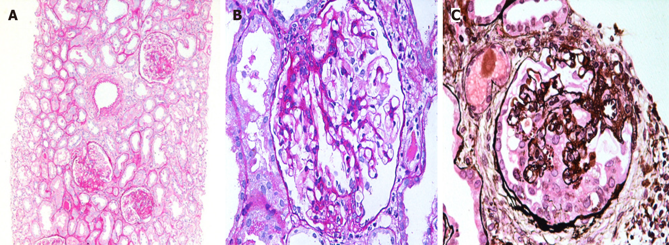
Figure 1 Histological features of common variants of focal segmental glomerulosclerosis.
A: Medium-power view showing three glomeruli with segmental scars in indeterminate locations in a case of focal segmental glomerulosclerosis (FSGS), not otherwise specified. Mild patchy tubular atrophy and interstitial scarring is seen in the background [Periodic Acid-Schiff (PAS), × 200]; B: High-power view showing one glomerulus with segmental scar and adhesion formation involving the tip domain diagonally opposite the vascular pole, an example of the TIP variant (PAS, × 400); C: High-power view showing one glomerulus with segmental collapse of capillary tufts accompanied by podocyte hypertrophy and hyperplasia in a case of collapsing FSGS. Many proteinaceous droplets are also seen in the cytoplasm of podocytes (Jone’s methenamine silver, × 400).
- Citation: Jafry NH, Manan S, Rashid R, Mubarak M. Clinicopathological features and medium-term outcomes of histologic variants of primary focal segmental glomerulosclerosis in adults: A retrospective study. World J Nephrol 2024; 13(1): 88028
- URL: https://www.wjgnet.com/2220-6124/full/v13/i1/88028.htm
- DOI: https://dx.doi.org/10.5527/wjn.v13.i1.88028


