Copyright
©The Author(s) 2023.
World J Nephrol. Sep 25, 2023; 12(4): 93-103
Published online Sep 25, 2023. doi: 10.5527/wjn.v12.i4.93
Published online Sep 25, 2023. doi: 10.5527/wjn.v12.i4.93
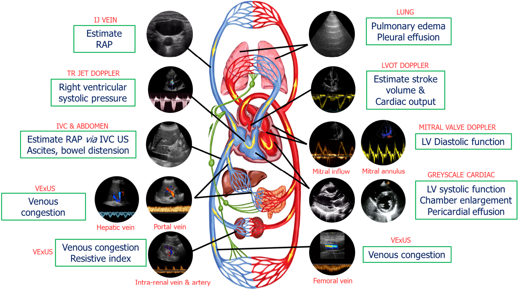
Figure 1 Common sonographic applications used by trained clinicians at the bedside in the evaluation of acute kidney injury.
It is important to evaluate multiple points of the hemodynamic circuit instead of relying on isolated parameters. IJ: Internal jugular; RAP: Right atrial pressure; TR: Tricuspid regurgitation; IVC: Inferior vena cava; US: Ultrasound; VExUS: venous excess ultrasound; LV: Left ventricular; LVOT: Left ventricular outflow tract. Citation: Koratala A, Reisinger N. Point of Care Ultrasound in Cirrhosis-Associated Acute Kidney Injury: Beyond Inferior Vena Cava. Kidney360 2022; 3: 1965-1968. Copyright© 2022 by the American Society of Nephrology (corresponding author’s prior open access publication).
- Citation: Batool A, Chaudhry S, Koratala A. Transcending boundaries: Unleashing the potential of multi-organ point-of-care ultrasound in acute kidney injury. World J Nephrol 2023; 12(4): 93-103
- URL: https://www.wjgnet.com/2220-6124/full/v12/i4/93.htm
- DOI: https://dx.doi.org/10.5527/wjn.v12.i4.93


