Copyright
©The Author(s) 2023.
World J Nephrol. Mar 25, 2023; 12(2): 29-39
Published online Mar 25, 2023. doi: 10.5527/wjn.v12.i2.29
Published online Mar 25, 2023. doi: 10.5527/wjn.v12.i2.29
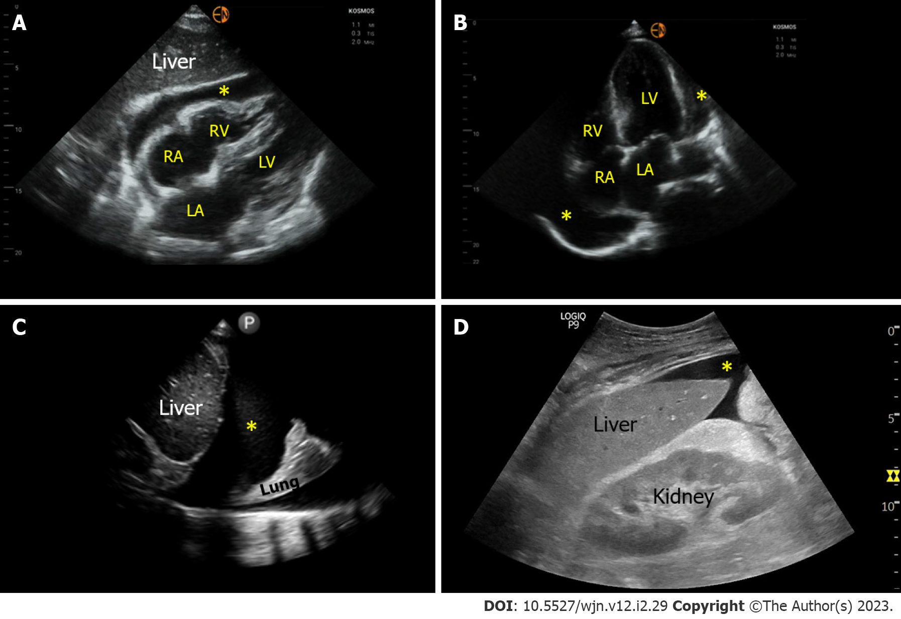
Figure 5 Sonographic images demonstrating various effusions.
A and B: Pericardial effusion (*) as seen on subxiphoid and apical 4-chamber cardiac views respectively; C: Right pleural effusion (*) seen from the lateral scanning window; D: Ascites (*) seen from the right upper quadrant. RA: Right atrium; LA: Left atrium; RV: Right ventricle; LV: Left ventricle.
- Citation: Bhasin-Chhabra B, Koratala A. Point of care ultrasonography in onco-nephrology: A stride toward better physical examination. World J Nephrol 2023; 12(2): 29-39
- URL: https://www.wjgnet.com/2220-6124/full/v12/i2/29.htm
- DOI: https://dx.doi.org/10.5527/wjn.v12.i2.29


