Copyright
©The Author(s) 2023.
World J Nephrol. Mar 25, 2023; 12(2): 29-39
Published online Mar 25, 2023. doi: 10.5527/wjn.v12.i2.29
Published online Mar 25, 2023. doi: 10.5527/wjn.v12.i2.29
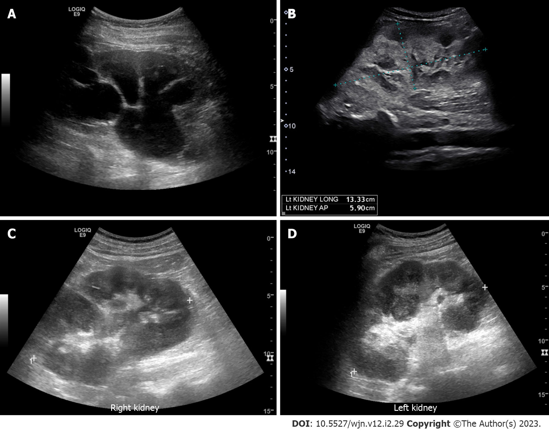
Figure 2 Renal ultrasound images demonstrating.
A: Severe hydronephrosis (branching anechoic area); B: Enlarged kidney with hyperechoic cortex in a patient with myeloma; C and D: Bilateral renal involvement with lymphoma. Note irregular outline and heterogenous parenchyma.
- Citation: Bhasin-Chhabra B, Koratala A. Point of care ultrasonography in onco-nephrology: A stride toward better physical examination. World J Nephrol 2023; 12(2): 29-39
- URL: https://www.wjgnet.com/2220-6124/full/v12/i2/29.htm
- DOI: https://dx.doi.org/10.5527/wjn.v12.i2.29


