Copyright
©2012 Baishideng.
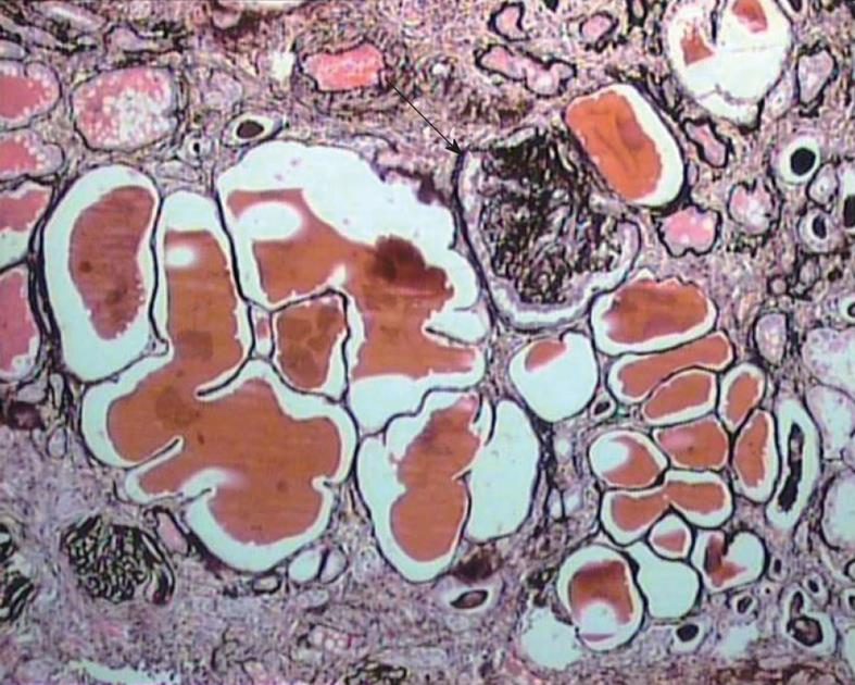
Figure 2 Medium-power view showing marked tubular atrophy associated with interstitial fibrosis and mononuclear inflammatory cell infiltrate in the interstitium.
Many dilated tubules contain eosinophilic casts. One glomerulus shows ischemic solidification and another one, global collapse with podocyter hypertrophy but little podocyte proliferation (arrow). Jones’ methenamine silver, × 200.
- Citation: Mubarak M. Collapsing focal segmental glomerulosclerosis: Current concepts. World J Nephrol 2012; 1(2): 35-42
- URL: https://www.wjgnet.com/2220-6124/full/v1/i2/35.htm
- DOI: https://dx.doi.org/10.5527/wjn.v1.i2.35


