Copyright
©2012 Baishideng.
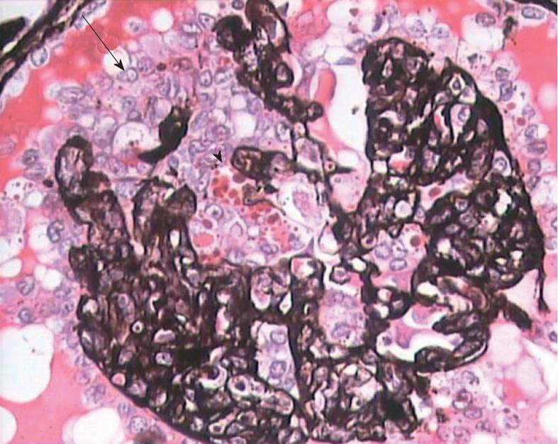
Figure 1 High magnification view showing global collapse of capillary tufts associated with marked hypertrophy and hyperplasia of podocytes (arrow), forming a pseudo-crescent.
Marked cytoplasmic vacuolization and protein resorption droplets (arrowhead) are seen in podocytes. Jones’ methenamine silver, × 400.
- Citation: Mubarak M. Collapsing focal segmental glomerulosclerosis: Current concepts. World J Nephrol 2012; 1(2): 35-42
- URL: https://www.wjgnet.com/2220-6124/full/v1/i2/35.htm
- DOI: https://dx.doi.org/10.5527/wjn.v1.i2.35


