Copyright
©The Author(s) 2017.
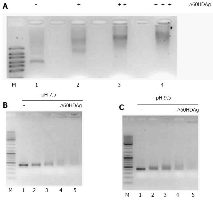
Figure 6 Gel retardation assay.
A: Binding of ∆60HDAg to HDV RNA. Purified recombinant ∆60HDAg was incubated, in standard pH 7.5 binding buffer, with 100 ng of HDV RNA at increasing concentrations (0, 0.5, 1.5, and 3 μmol/L, respectively). Left in each panel is a RNA marker (RiboRuler High Range RNA Ladder, Fermentas); B and C: ∆60HDAg binding to DNA and HDV RNA, respectively. In panel B, 100 ng of dsDNA were incubated in standard pH 7.5 binding buffer, with increasing concentrations of purified recombinant ∆60HDAg (0, 2, 4, 6, 8, 10, and 12 μmol/L). Panel C shows the assay in binding buffer at pH 9.5. Recombinant ∆60HDAg was incubated with 100 ng of dsDNA, at different concentrations (0, 2, 4, 6, 8, 10, and 12 μmol/L). In panel C, 100 ng of HDV RNA was incubated, in standard pH 9.5 binding buffer, with increasing concentrations of purified recombinant ∆60HDAg (0, 0.5, 1, 1.5, and 2 μmol/L). HDV: Hepatitis delta virus.
- Citation: Alves C, Cheng H, Tavanez JP, Casaca A, Gudima S, Roder H, Cunha C. Structural and nucleic acid binding properties of hepatitis delta virus small antigen. World J Virol 2017; 6(2): 26-35
- URL: https://www.wjgnet.com/2220-3249/full/v6/i2/26.htm
- DOI: https://dx.doi.org/10.5501/wjv.v6.i2.26


