Copyright
©The Author(s) 2016.
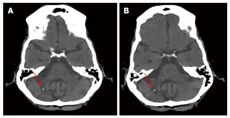
Figure 1 Computed tomography scans show no progression.
A: Axial slice from the CT scan of 2006 showing the cerebellar lesion (red arrow), a small calcification, and atherosclerosis of cerebral vasculature; B: Axial slice of the same coordinates as in (A) from the CT scan of 2008 showing the cerebellar lesion (red arrow), as well as the calcification. Note the radiolucency of the cerebellar peduncle and the loss of tissue density in the cerebellum. This location corresponds to that of the 1999 biopsy and the histologic sections examined at autopsy. CT: Computed tomography.
- Citation: SantaCruz KS, Roy G, Spigel J, Bearer EL. Neuropathology of JC virus infection in progressive multifocal leukoencephalopathy in remission. World J Virol 2016; 5(1): 31-37
- URL: https://www.wjgnet.com/2220-3249/full/v5/i1/31.htm
- DOI: https://dx.doi.org/10.5501/wjv.v5.i1.31


