Copyright
©2013 Baishideng Publishing Group Co.
World J Virol. Aug 12, 2013; 2(3): 123-135
Published online Aug 12, 2013. doi: 10.5501/wjv.v2.i3.123
Published online Aug 12, 2013. doi: 10.5501/wjv.v2.i3.123
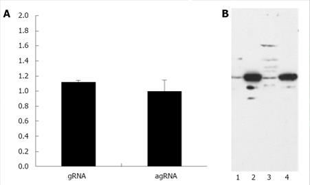
Figure 2 Nucleo-cytoplasmic distribution of hepatitis delta virus gRNA and agRNA.
HuH-7 cells were trasnfected with plasmids pDL542 and pDL481, respectively. A: The relative quantification of HDV RNA was performed by real time-polymerase chain reaction using the 2-∆∆Ct method. Results are presented as the cytoplasmic /nuclear ratio (C/N) and correspond to the mean of three independent experiments. Bars indicate the standard deviation; B: Western blotting analysis of nuclear and cytoplasmic HuH-7 cell protein fractions. Equivalent amounts of nuclear (lanes 1 and 3) and cytoplasmic (lanes 2 and 4) protein fractions used for quantification of gRNA (lanes 1 and 2) and agRNA (lanes 3 and 4) were separated in 12% SDS-PAGE gels. The possible contamination of nuclear fractions was monitored by using an anti-GAPDH antibody.
- Citation: Freitas N, Cunha C. Searching for nuclear export elements in hepatitis D virus RNA. World J Virol 2013; 2(3): 123-135
- URL: https://www.wjgnet.com/2220-3249/full/v2/i3/123.htm
- DOI: https://dx.doi.org/10.5501/wjv.v2.i3.123


