Copyright
©The Author(s) 2022.
World J Virol. Sep 25, 2022; 11(5): 331-340
Published online Sep 25, 2022. doi: 10.5501/wjv.v11.i5.331
Published online Sep 25, 2022. doi: 10.5501/wjv.v11.i5.331
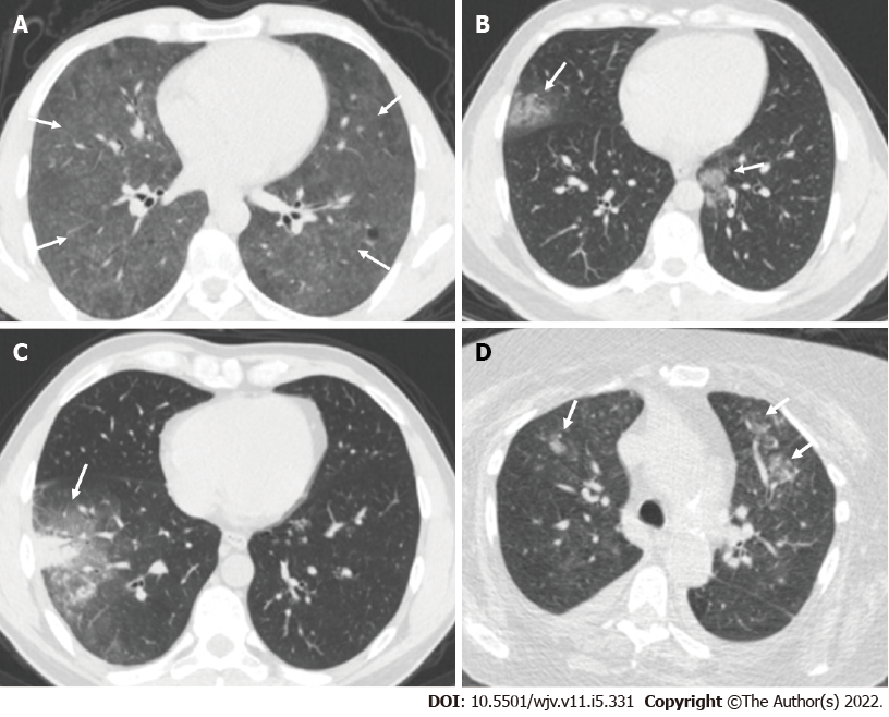
Figure 5 Thoracic computed tomography scans in patients.
A: A 45-year-old male patient underwent a thoracic computed tomography (CT) scan. Sections passing through the middle zones showed diffuse ground-glass infiltration areas with preservation of subpleural areas in both lungs (arrows); B: A 44-year-old female patient underwent a thoracic CT scan. In the sections passing through the lower zones, infiltration areas of peripheral ground glass density with a mild halo were observed in the medial basal segment on the right and in the upper lobe inferior lingular segment on the left (arrows); C: A 35-year-old male patient underwent a thoracic CT scan. A subpleural consolidation area with air bronchogram and ground-glass density halo could be seen in the lateral basal segment of the right lung lower lobe in sections passing through the lower zones (arrow); D: A 77-year-old female patient underwent a thoracic CT scan. In the sections passing through the upper zones, centrilobular nodular infiltrating areas in the form of a budded tree pattern were observed in the anterior segments of the upper lobes of bilateral lungs, particularly on the left (arrows).
- Citation: Karavas E, Unver E, Aydın S, Yalcin GS, Fatihoglu E, Kuyrukluyildiz U, Arslan YK, Yazici M. Effect of age on computed tomography findings: Specificity and sensitivity in coronavirus disease 2019 infection. World J Virol 2022; 11(5): 331-340
- URL: https://www.wjgnet.com/2220-3249/full/v11/i5/331.htm
- DOI: https://dx.doi.org/10.5501/wjv.v11.i5.331


