Copyright
©The Author(s) 2017.
World J Transplant. Aug 24, 2017; 7(4): 243-249
Published online Aug 24, 2017. doi: 10.5500/wjt.v7.i4.243
Published online Aug 24, 2017. doi: 10.5500/wjt.v7.i4.243
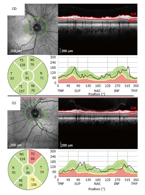
Figure 5 Optical coherence tomography showed a localized defect in the temporal-superior area of the peripapillary retinal nerve fiber layer of the left eye (OS).
In the right eye (OD), the retinal nerve fiber layer thickness was normal in all peripapillary locations.
- Citation: Gama IF, Almeida LD. De novo intraocular amyloid deposition after hepatic transplantation in familial amyloidotic polyneuropathy. World J Transplant 2017; 7(4): 243-249
- URL: https://www.wjgnet.com/2220-3230/full/v7/i4/243.htm
- DOI: https://dx.doi.org/10.5500/wjt.v7.i4.243


