Copyright
©The Author(s) 2017.
World J Transplant. Aug 24, 2017; 7(4): 243-249
Published online Aug 24, 2017. doi: 10.5500/wjt.v7.i4.243
Published online Aug 24, 2017. doi: 10.5500/wjt.v7.i4.243
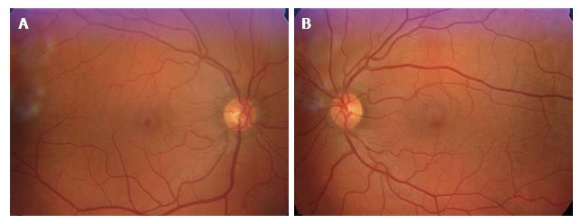
Figure 3 Retinography of right eye (A) and left eye (B) showed normal posterior poles, which were clearly visible due to the mild amyloid deposition in the vitreous, with only few opacities, which did not compromise visual acuity.
- Citation: Gama IF, Almeida LD. De novo intraocular amyloid deposition after hepatic transplantation in familial amyloidotic polyneuropathy. World J Transplant 2017; 7(4): 243-249
- URL: https://www.wjgnet.com/2220-3230/full/v7/i4/243.htm
- DOI: https://dx.doi.org/10.5500/wjt.v7.i4.243


