Copyright
©The Author(s) 2017.
World J Transplant. Aug 24, 2017; 7(4): 243-249
Published online Aug 24, 2017. doi: 10.5500/wjt.v7.i4.243
Published online Aug 24, 2017. doi: 10.5500/wjt.v7.i4.243
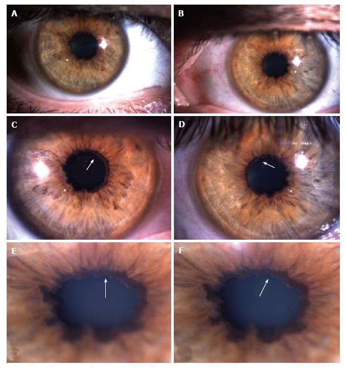
Figure 1 Slit-lamp photos showing scalloped pupils (astherisks) and amyloid deposition in the pupillary border (arrows) in both eyes.
A and B: Slit-lamp photos of anterior segment of right (A) and left eyes (B) at low magnification; C and D: Slit-lamp photos at higher magnification to show pupillary margins of right (C) and left (D) eyes with more detail, in order to highlight the irregular pupillary margins, the scalloped pupils (astherisks) with amyloid deposits (arrows); E and F: Slit-lamp photos of the right eye (E and F) at the highest magnification to enhance visualization of the pupillary amyloid deposits (arrows), which resemble those seen in pseudoexfoliation syndrome.
- Citation: Gama IF, Almeida LD. De novo intraocular amyloid deposition after hepatic transplantation in familial amyloidotic polyneuropathy. World J Transplant 2017; 7(4): 243-249
- URL: https://www.wjgnet.com/2220-3230/full/v7/i4/243.htm
- DOI: https://dx.doi.org/10.5500/wjt.v7.i4.243


