Copyright
©The Author(s) 2017.
World J Transplant. Feb 24, 2017; 7(1): 98-102
Published online Feb 24, 2017. doi: 10.5500/wjt.v7.i1.98
Published online Feb 24, 2017. doi: 10.5500/wjt.v7.i1.98
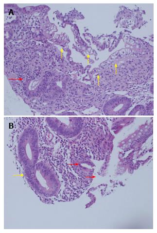
Figure 3 Small bowel allograft biopsy - day 23.
A: Mucosal erosion with marked surface enterocyte degeneration and cytoplasmic vacuolation, sloughing (yellow arrows), inflamed granulation-like tissue within the lamina propria, prominent crypt injury (red arrow) and focal drop out; B: Cryptitis with increased epithelial apoptosis (yellow arrow), mixed lamina propria inflammatory infiltrate and surface epithelial erosion (red arrows).
- Citation: Apostolov R, Asadi K, Lokan J, Kam N, Testro A. Mycophenolate mofetil toxicity mimicking acute cellular rejection in a small intestinal transplant. World J Transplant 2017; 7(1): 98-102
- URL: https://www.wjgnet.com/2220-3230/full/v7/i1/98.htm
- DOI: https://dx.doi.org/10.5500/wjt.v7.i1.98


