Copyright
©The Author(s) 2017.
World J Transplant. Feb 24, 2017; 7(1): 88-93
Published online Feb 24, 2017. doi: 10.5500/wjt.v7.i1.88
Published online Feb 24, 2017. doi: 10.5500/wjt.v7.i1.88
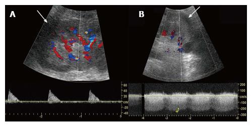
Figure 2 Presence of peri-allograft hematoma and Doppler ultrasound findings.
A: Transplant arterial flow. Peri-allograft hypoechoic area (arrows) with absent diastolic flow in the arcuate arteries; B: Transplant venous flow. The transplant renal vein was patent.
- Citation: Takahashi K, Prashar R, Putchakayala KG, Kane WJ, Denny JE, Kim DY, Malinzak LE. Allograft loss from acute Page kidney secondary to trauma after kidney transplantation. World J Transplant 2017; 7(1): 88-93
- URL: https://www.wjgnet.com/2220-3230/full/v7/i1/88.htm
- DOI: https://dx.doi.org/10.5500/wjt.v7.i1.88


