Copyright
©The Author(s) 2017.
World J Transplant. Feb 24, 2017; 7(1): 43-48
Published online Feb 24, 2017. doi: 10.5500/wjt.v7.i1.43
Published online Feb 24, 2017. doi: 10.5500/wjt.v7.i1.43
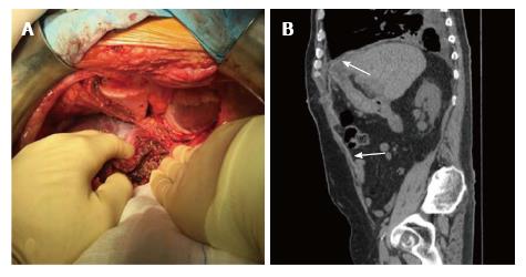
Figure 3 The abdominal exploration showed a neoplasm of left lobe liver graft with infiltration of the diaphragm which extended to the pleura and pericardium.
A: Left liver lobectomy of the graft with resection of the diaphragm “en bloc” with adjacent portion of right pleura and pericardium; B: Computed tomography scan at 6 mo after abdominal wall repair (arrow: Biological prosthesis).
- Citation: Vennarecci G, Mascianà G, De Werra E, Sandri GBL, Ferraro D, Burocchi M, Tortorelli G, Guglielmo N, Ettorre GM. Effectiveness and versatility of biological prosthesis in transplanted patients. World J Transplant 2017; 7(1): 43-48
- URL: https://www.wjgnet.com/2220-3230/full/v7/i1/43.htm
- DOI: https://dx.doi.org/10.5500/wjt.v7.i1.43


