Copyright
©The Author(s) 2017.
World J Transplant. Feb 24, 2017; 7(1): 1-25
Published online Feb 24, 2017. doi: 10.5500/wjt.v7.i1.1
Published online Feb 24, 2017. doi: 10.5500/wjt.v7.i1.1
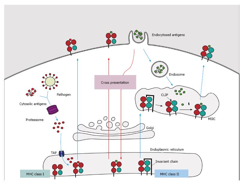
Figure 1 Major histocompatibility complex class I and II pathways.
(1) MHC class I molecules present peptides derived from proteins presented in the cytosol of endogenous or pathogen origin. The proteasome breaks down these proteins into peptides, which are then translocated to ER by the transporter associated with antigen processing (TAP) to access the MHC class I molecules. In absence of peptides, MHC class I molecule is stabilized by ER chaperones (calreticulin, PDIA3, PDI and tapasin), but when peptides with sufficient affinity bind to class I molecules, these chaperones are released and the peptide: MHC complex leaves the ER for presentation on cell surface of CD8+ T cells; (2) MHC class II molecules present peptides derived from proteins that enter the cell through endocytosis. The chains α and β are assembled in the endoplasmatic reticulum associated with the invariant-chain (li) to prevent binding of endogenous proteins. This complex (MHC:li) is translocated to MHC class II compartment (MIIC) where li is degraded to class II-associated invariant chain (CLIP). In the MIIC the MHC class II molecules acquire HLA-DM to facilitate the exchange of CLIP to specific antigen derived from degraded protein on the endosomal pathway, thus the complexes are transported to the plasma membrane to present the peptide to CD4+ T cells; (3) Cross presentation involves dendritic cells with the unique ability to present exogenous antigens via MHC class I (by a mechanism not completely understood). MHC: Major histocompatibility complex.
- Citation: da Silva MB, da Cunha FF, Terra FF, Camara NOS. Old game, new players: Linking classical theories to new trends in transplant immunology. World J Transplant 2017; 7(1): 1-25
- URL: https://www.wjgnet.com/2220-3230/full/v7/i1/1.htm
- DOI: https://dx.doi.org/10.5500/wjt.v7.i1.1


