Copyright
©The Author(s) 2016.
World J Transplant. Jun 24, 2016; 6(2): 278-290
Published online Jun 24, 2016. doi: 10.5500/wjt.v6.i2.278
Published online Jun 24, 2016. doi: 10.5500/wjt.v6.i2.278
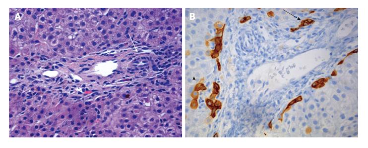
Figure 4 Progressive familial intrahepatic cholestasis type 1 and 2 can also present with duct paucity.
A and B: The portal tracts show an absence of bile duct with periportal duct reaction; B: A higher power view of the portal tract with vein on the left artery on the right (arrow) and no appreciable bile duct. Keratin 7 is negative in this portal tract in B and positive in the bile duct reaction (arrow) with some bile duct progenitor cells (paler brown staining arrowhead).
- Citation: Mehl A, Bohorquez H, Serrano MS, Galliano G, Reichman TW. Liver transplantation and the management of progressive familial intrahepatic cholestasis in children. World J Transplant 2016; 6(2): 278-290
- URL: https://www.wjgnet.com/2220-3230/full/v6/i2/278.htm
- DOI: https://dx.doi.org/10.5500/wjt.v6.i2.278


