Copyright
©The Author(s) 2015.
World J Transplant. Dec 24, 2015; 5(4): 360-365
Published online Dec 24, 2015. doi: 10.5500/wjt.v5.i4.360
Published online Dec 24, 2015. doi: 10.5500/wjt.v5.i4.360
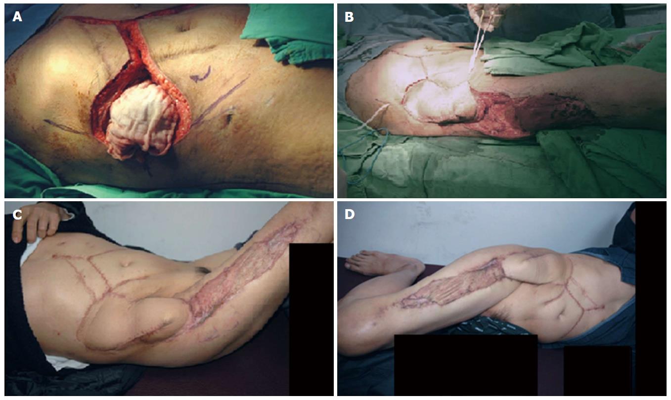
Figure 3 Recipient’s intraoperative and follow up images.
A: Ten centimeter × 10 cm diameter right abdominal wall defect following the wide local excision of the area; B: Perioperative picture of the transposition of the right thigh extended pedicle flap and coverage of the right side abdominal wall defect; C: Post operative picture from the outpatient clinic on a three months follow up; D: Post operative picture from the outpatient clinic on a six months follow up.
- Citation: Yang HR, Thorat A, Gesakis K, Li PC, Kiranantawat K, Chen HC, Jeng LB. Living donor liver transplantation with abdominal wall reconstruction for hepatocellular carcinoma with needle track seeding. World J Transplant 2015; 5(4): 360-365
- URL: https://www.wjgnet.com/2220-3230/full/v5/i4/360.htm
- DOI: https://dx.doi.org/10.5500/wjt.v5.i4.360


