Copyright
©The Author(s) 2015.
World J Transplant. Dec 24, 2015; 5(4): 360-365
Published online Dec 24, 2015. doi: 10.5500/wjt.v5.i4.360
Published online Dec 24, 2015. doi: 10.5500/wjt.v5.i4.360
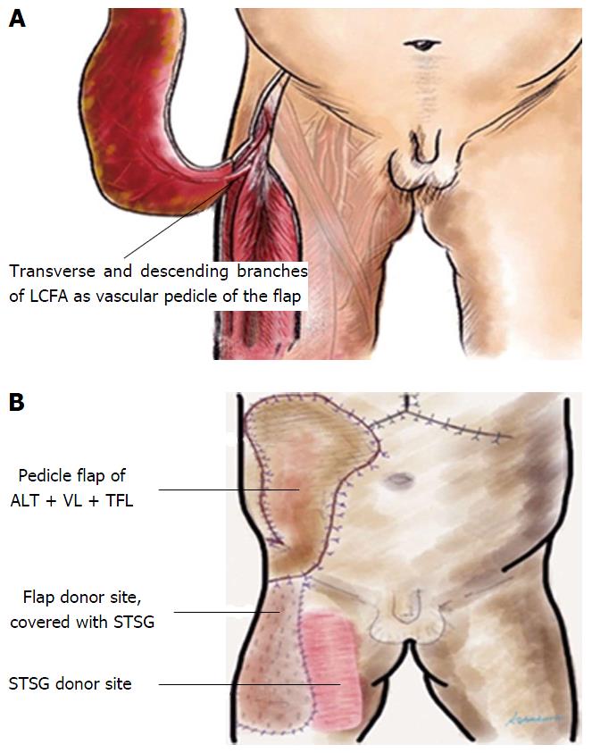
Figure 2 Diagramtic depiction of the myocutaneous pedicle flap for abdominal wall reconstruction.
A: Extended right thigh flap based on the transverse and descending branches of the LCFA; B: Pedicle flap of ALT + VL + TFL for coverage of right abdominal wall defect. Donor site covered with STSG taken from the right thigh. LCFA: Lateral circumflex femoral artery; ALT: Anterolateral thigh; VL: Vastus lateralis; TFL: Tensor fascia latae; STSG: Split thickness skin graft.
- Citation: Yang HR, Thorat A, Gesakis K, Li PC, Kiranantawat K, Chen HC, Jeng LB. Living donor liver transplantation with abdominal wall reconstruction for hepatocellular carcinoma with needle track seeding. World J Transplant 2015; 5(4): 360-365
- URL: https://www.wjgnet.com/2220-3230/full/v5/i4/360.htm
- DOI: https://dx.doi.org/10.5500/wjt.v5.i4.360


