Copyright
©The Author(s) 2015.
World J Transplant. Dec 24, 2015; 5(4): 354-359
Published online Dec 24, 2015. doi: 10.5500/wjt.v5.i4.354
Published online Dec 24, 2015. doi: 10.5500/wjt.v5.i4.354
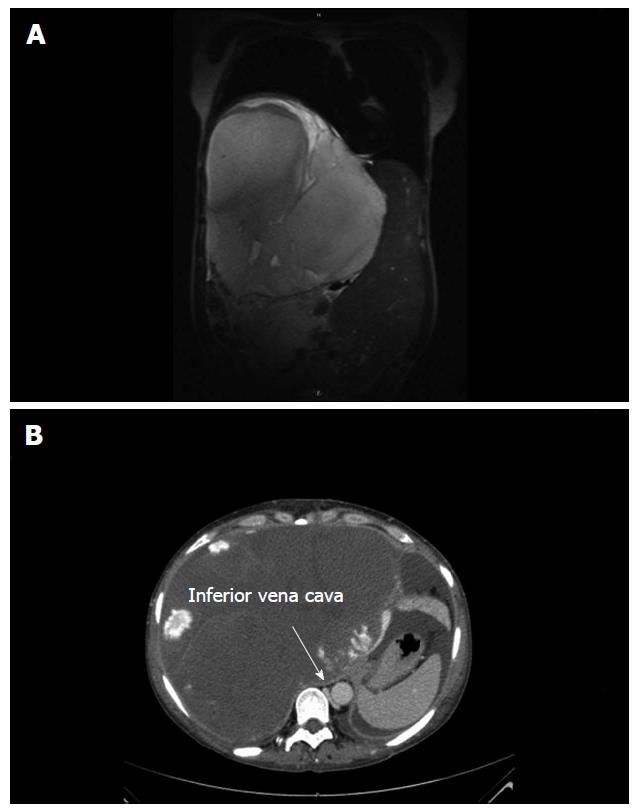
Figure 1 Radiological imaging.
A: Radiological imaging showing a tumour of 21.7 cm × 23.7 cm × 25.5 cm in size in segments V-VIII. The tumour volume was 6000 mL; the total liver volume was calculated as 9691 mL; B: The vena cava inferior was massively dislocated to the left and slit-shaped due to compression. This contributes to progressive ascites.
- Citation: Lange UG, Bucher JN, Schoenberg MB, Benzing C, Schmelzle M, Gradistanac T, Strocka S, Hau HM, Bartels M. Orthotopic liver transplantation for giant liver haemangioma: A case report. World J Transplant 2015; 5(4): 354-359
- URL: https://www.wjgnet.com/2220-3230/full/v5/i4/354.htm
- DOI: https://dx.doi.org/10.5500/wjt.v5.i4.354


