Copyright
©The Author(s) 2025.
World J Transplant. Jun 18, 2025; 15(2): 98620
Published online Jun 18, 2025. doi: 10.5500/wjt.v15.i2.98620
Published online Jun 18, 2025. doi: 10.5500/wjt.v15.i2.98620
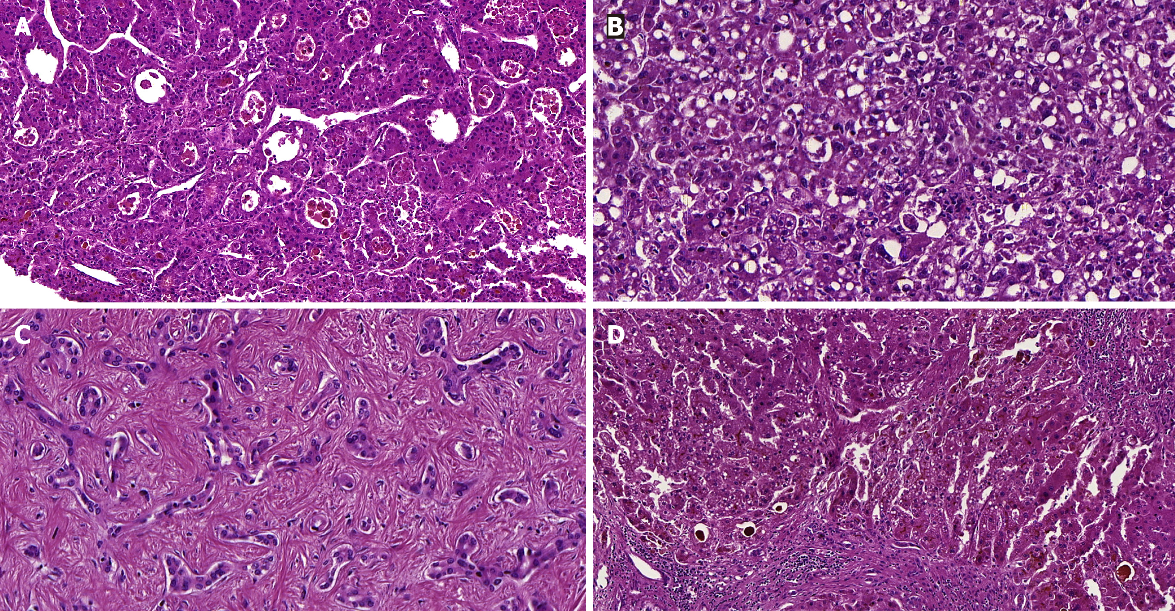
Figure 2 Microscopic characteristics of the liver explant, with advanced chronic disease and neoplastic nodules.
A: This hepatic nodule displays a moderately-differentiated hepatocellular carcinoma (HCC) with many pseudoglands and bile pigment in dilated canaliculi [100×, hematoxylin and eosin (HE) stain]; B: Features of the steatohepatitic subtype of HCC were seen in some areas of the nodules, such as ballooned hepatocytes, Mallory-Denk bodies and thin fibrous bands (200×, HE stain); C: A very well-differentiated adenocarcinoma with ductular configuration (cholangiolocarcinoma) was detected in a nodule, with small-tubular and cord-like formations, embedded in a fibrous stroma (200×, HE stain); D: The cirrhotic liver demonstrated areas with marked cholestasis probably related to the acute clinical deterioration, as well as steatosis and other findings (100×, HE stain).
- Citation: Kasputis Zanini LY, Lima FR, Fernandes MR, Alvarez PSE, Silva MS, Martins Filho APR, Franzini TAP, Nacif LS. Ischemic colitis with small-vessel occlusion, simultaneous total colectomy and liver transplantation: A case report. World J Transplant 2025; 15(2): 98620
- URL: https://www.wjgnet.com/2220-3230/full/v15/i2/98620.htm
- DOI: https://dx.doi.org/10.5500/wjt.v15.i2.98620


