Copyright
©The Author(s) 2025.
World J Transplant. Jun 18, 2025; 15(2): 98620
Published online Jun 18, 2025. doi: 10.5500/wjt.v15.i2.98620
Published online Jun 18, 2025. doi: 10.5500/wjt.v15.i2.98620
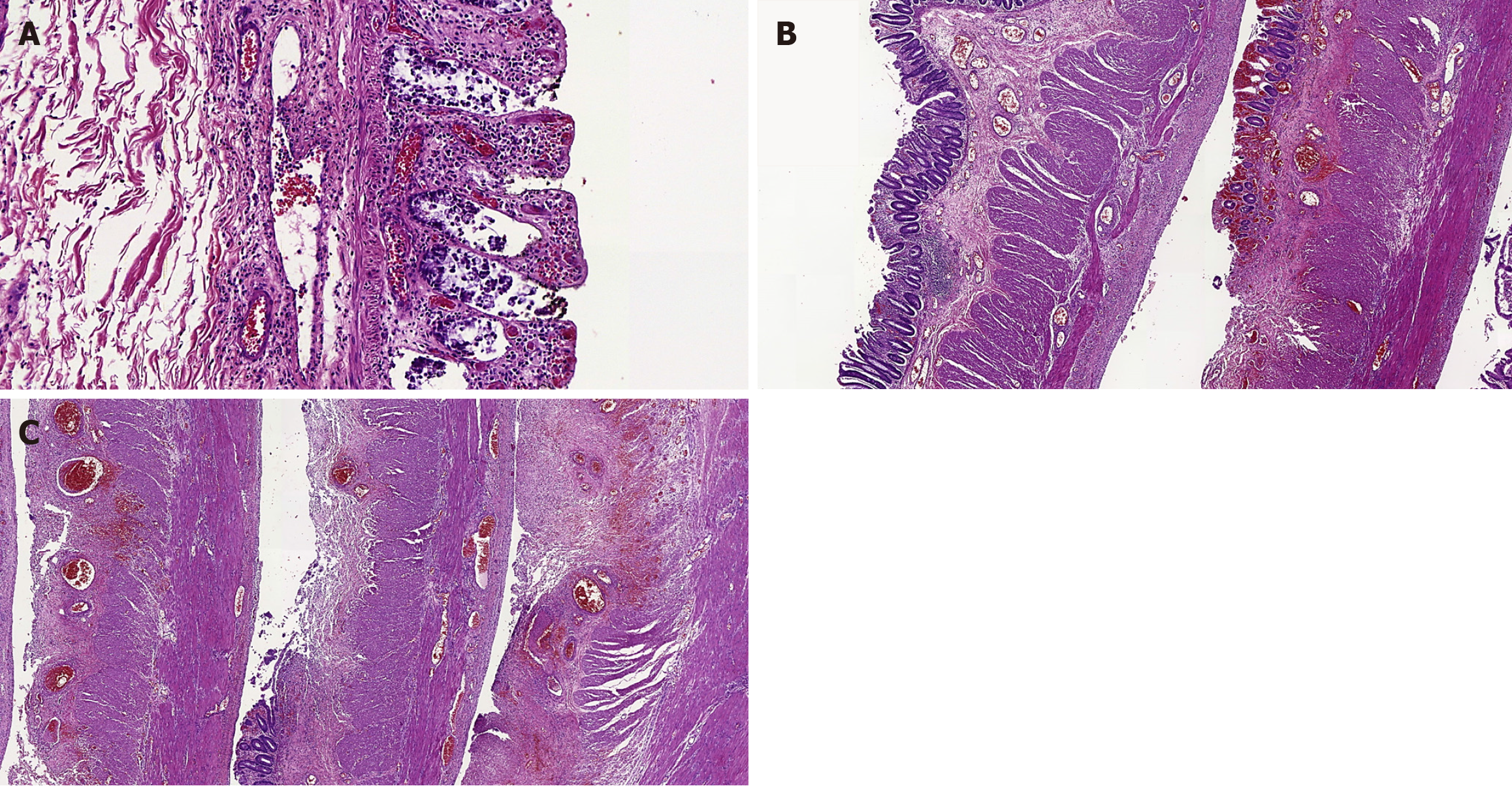
Figure 1 Histopathological alterations in the resected colon with patchy ischemic features.
A: The colonic mucosa shows superficial necrosis and some fibrin thrombi in small vessels of the lamina propria [100×, hematoxylin and eosin (HE) stain]; B: In this field, one of the intestinal fragments has signs of hemorrhage, congestion and an area of ulceration, whereas the other depicts only subtle alterations in the mucosa (20×, HE stain); C: This picture reveals necrosis and ulcerations in all colonic fragments, as well as a pseudomembrane in one of them and congested vessels in the wall (20×, HE stain).
- Citation: Kasputis Zanini LY, Lima FR, Fernandes MR, Alvarez PSE, Silva MS, Martins Filho APR, Franzini TAP, Nacif LS. Ischemic colitis with small-vessel occlusion, simultaneous total colectomy and liver transplantation: A case report. World J Transplant 2025; 15(2): 98620
- URL: https://www.wjgnet.com/2220-3230/full/v15/i2/98620.htm
- DOI: https://dx.doi.org/10.5500/wjt.v15.i2.98620


