Copyright
©The Author(s) 2025.
World J Transplant. Jun 18, 2025; 15(2): 100373
Published online Jun 18, 2025. doi: 10.5500/wjt.v15.i2.100373
Published online Jun 18, 2025. doi: 10.5500/wjt.v15.i2.100373
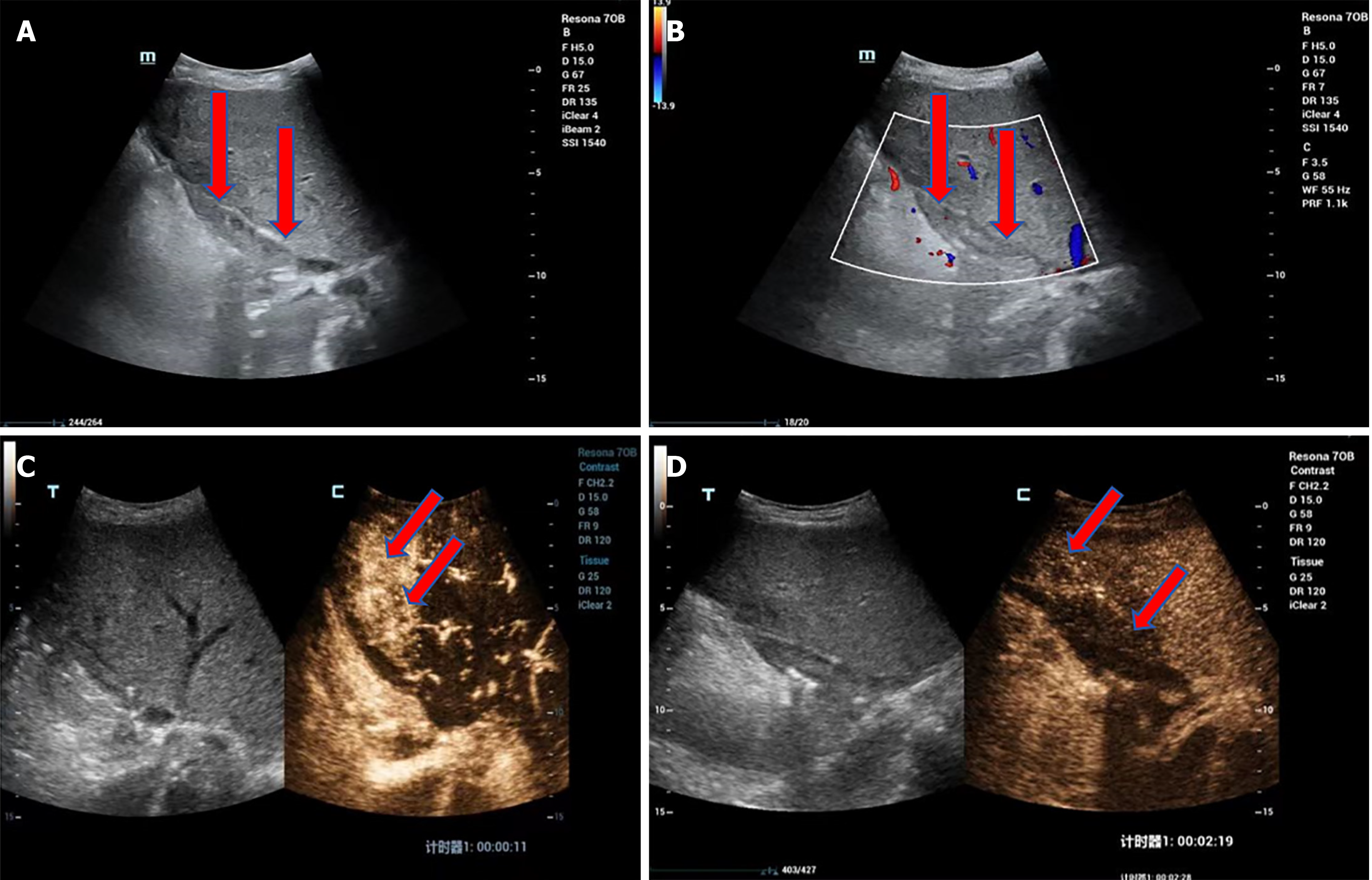
Figure 4 Bridging vein thrombosis in a 37-year-old female with primary sclerosing cholangitis and decompensated cirrhosis.
A: Grayscale ultrasound shows the bridging vein lumen filled with medium to low echogenic material; B: Color Doppler ultrasound shows no significant blood flow signal within the bridging vein lumen, suggesting occlusion due to bridging vein thrombosis; C: Contrast-enhanced ultrasound (CEUS) shows no enhancement within the bridging vein lumen, confirming complete occlusion. An interesting phenomenon observed on CEUS is arterial phase hyperenhancement in the drainage area of the bridging vein (segment 4, indicated by the arrow); D: CEUS shows delayed phase hypoenhancement in the drainage area of the bridging vein (segment 4, indicated by the arrow). The abnormal arterial phase enhancement of the hepatic parenchyma in the drainage area is associated with local liver sinusoidal congestion due to impaired hepatic outflow. Surgical procedure: split liver transplantation (including left hemiliver with the middle hepatic vein). The donor liver was from a dextran-binding domain adult, split along the middle hepatic vein, with the left liver graft allocated to this patient. Intraoperative reconstruction of the proximal end of the middle hepatic vein was performed. Ultrasound examination, 1 month postoperatively. Biochemical indicators show elevated transaminases and bilirubin.
- Citation: Zhao NB, Luo Z, Li Y, Xia R, Zhang Y, Li YJ, Zhao D. Diagnostic value of ultrasonography for post-liver transplant hepatic vein complications. World J Transplant 2025; 15(2): 100373
- URL: https://www.wjgnet.com/2220-3230/full/v15/i2/100373.htm
- DOI: https://dx.doi.org/10.5500/wjt.v15.i2.100373


