Copyright
©The Author(s) 2025.
World J Transplant. Jun 18, 2025; 15(2): 100373
Published online Jun 18, 2025. doi: 10.5500/wjt.v15.i2.100373
Published online Jun 18, 2025. doi: 10.5500/wjt.v15.i2.100373
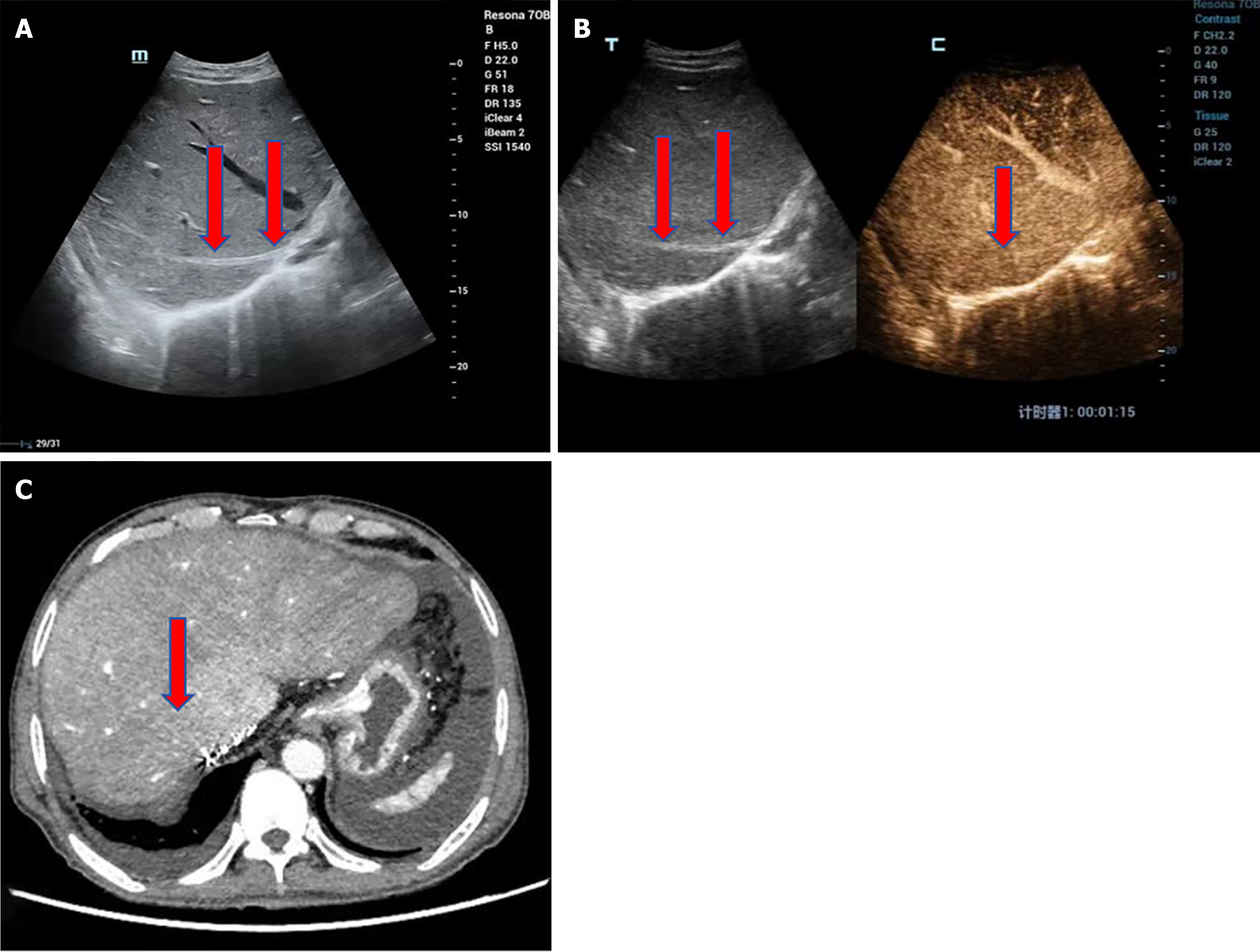
Figure 3 Hepatic vein occlusion in a 72-year-old male with decompensated hepatitis B cirrhosis.
A: The liver is significantly enlarged with coarse, dense parenchymal echotexture and patchy hypoechoic areas. The right hepatic vein appears narrowed with thickened, hyperechoic walls. Hepatic vein occlusion results in impaired hepatic blood outflow, leading to liver congestion and enlargement. Ultrasound can measure the maximum oblique diameter of the right lobe to assess liver size. One year post-transplantation, the liver size typically normalizes, with a right lobe maximum oblique diameter not exceeding 140 mm. In this patient, the liver is markedly enlarged. Chronic hepatic congestion can lead to increased parenchymal echogenicity and fibrosis, with patchy hypoechoic areas representing focal congestion; B: Contrast-enhanced ultrasound shows no enhancement within the right hepatic vein, confirming the diagnosis of right hepatic vein occlusion; C: Contrast-enhanced computed tomography reveals no enhancement in the right hepatic vein and heterogeneous liver density. Surgical procedure: Piggyback liver transplantation. Ultrasound examination: 14 years postoperatively. Clinical presentation: Massive ascites.
- Citation: Zhao NB, Luo Z, Li Y, Xia R, Zhang Y, Li YJ, Zhao D. Diagnostic value of ultrasonography for post-liver transplant hepatic vein complications. World J Transplant 2025; 15(2): 100373
- URL: https://www.wjgnet.com/2220-3230/full/v15/i2/100373.htm
- DOI: https://dx.doi.org/10.5500/wjt.v15.i2.100373


