Copyright
©The Author(s) 2025.
World J Transplant. Jun 18, 2025; 15(2): 100373
Published online Jun 18, 2025. doi: 10.5500/wjt.v15.i2.100373
Published online Jun 18, 2025. doi: 10.5500/wjt.v15.i2.100373
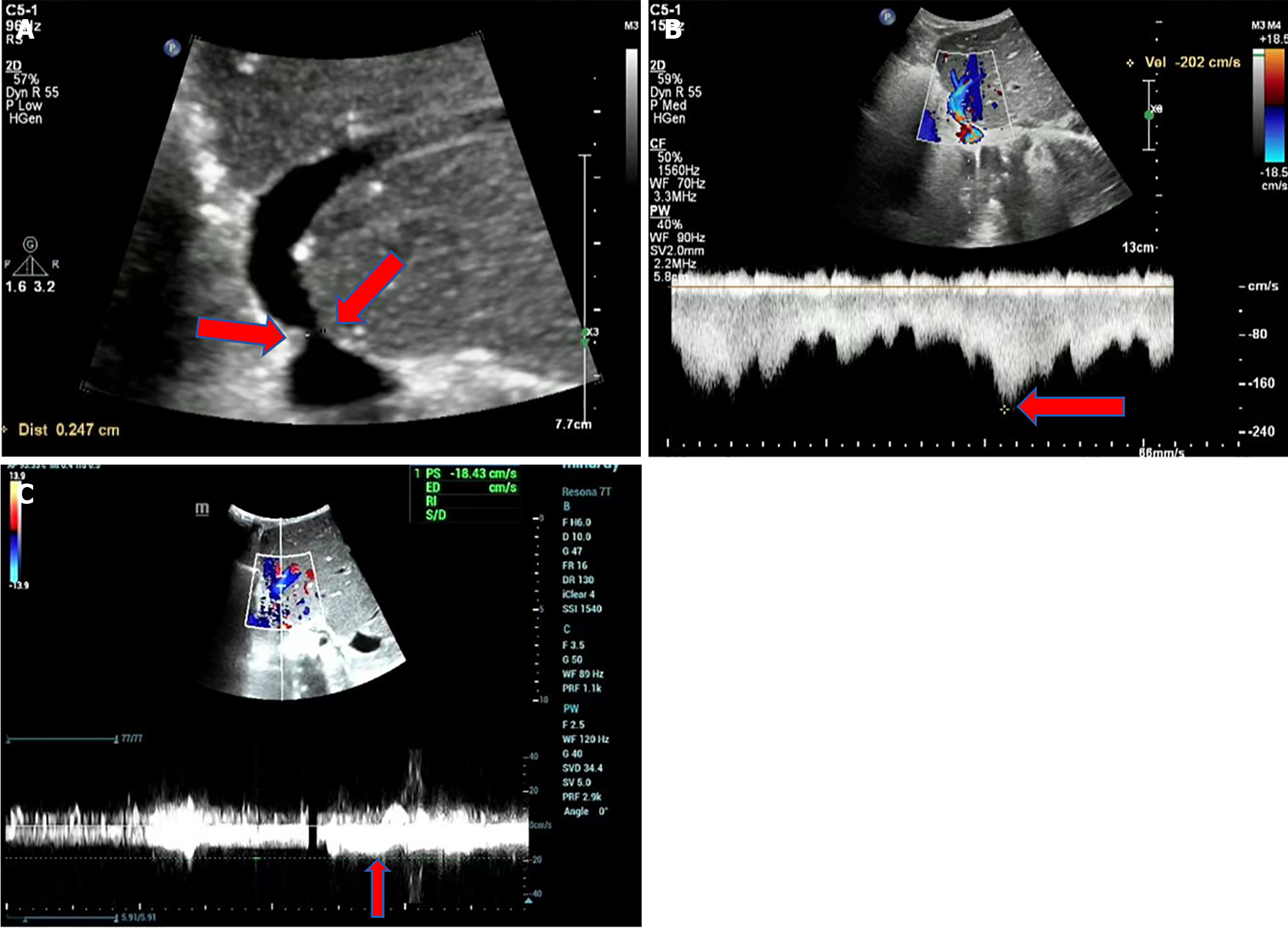
Figure 2 Hepatic vein stenosis in a 2-year-old girl with congenital biliary atresia.
A: Grayscale ultrasound shows a reduced diameter (2.4 mm) at the hepatic vein anastomosis site (red arrow). While grayscale ultrasound can reveal a narrowed diameter at the hepatic vein anastomosis site, the diagnosis of hepatic vein stenosis cannot be based solely on the reduced diameter; B: Color Doppler and spectral Doppler imaging demonstrate aliasing of blood flow signals and increased flow velocity (202 cm/second) at the hepatic vein anastomosis site; C: Spectral Doppler imaging shows decreased blood flow velocity in the main hepatic vein (18 cm/second). When the velocity ratio at the stenotic site to the pre-stenotic site exceeds 4:1, and there is a flattened hepatic vein waveform distal to the stenosis, hepatic vein outflow stenosis should be suspected. Surgical procedure: Left lobe living donor liver transplantation. Ultrasound examination: 2 years postoperatively. No abnormal clinical indicators.
- Citation: Zhao NB, Luo Z, Li Y, Xia R, Zhang Y, Li YJ, Zhao D. Diagnostic value of ultrasonography for post-liver transplant hepatic vein complications. World J Transplant 2025; 15(2): 100373
- URL: https://www.wjgnet.com/2220-3230/full/v15/i2/100373.htm
- DOI: https://dx.doi.org/10.5500/wjt.v15.i2.100373


