Copyright
©The Author(s) 2025.
World J Transplant. Mar 18, 2025; 15(1): 99220
Published online Mar 18, 2025. doi: 10.5500/wjt.v15.i1.99220
Published online Mar 18, 2025. doi: 10.5500/wjt.v15.i1.99220
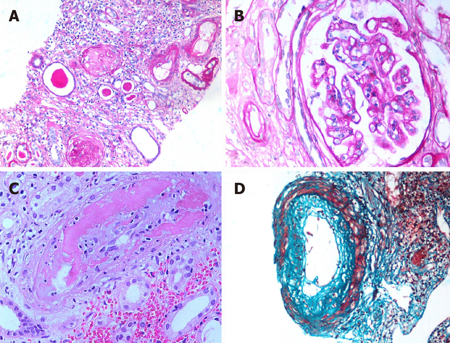
Figure 4 Pathology of acute and chronic antibody-mediated graft injury.
The principal target of antibodies is the microcirculation and larger blood vessels. A: Severe interstitial fibrosis and tubular atrophy. This lesion is entirely non-specific and may result from a variety of immune and non-immune causes [Periodic acid-Schiff (PAS) stain, × 200]; B: A glomerulus showing double contouring of capillary walls and an occasional inflammatory cell infiltration obliterating the capillary lumen (arrow) (PAS, × 400); C: A small artery exhibiting fibrinoid necrosis of the wall (v3). This lesion is strongly suggestive of active antibody-mediated rejection (AMR) (HE, × 400); D: Moderate fibrointimal thickening of an interlobular size artery. This lesion may be seen in chronic AMR as well as chronic T cell-mediated rejection (Trichrome stain, × 400).
- Citation: Abbas K, Mubarak M. Expanding role of antibodies in kidney transplantation. World J Transplant 2025; 15(1): 99220
- URL: https://www.wjgnet.com/2220-3230/full/v15/i1/99220.htm
- DOI: https://dx.doi.org/10.5500/wjt.v15.i1.99220


