Copyright
©The Author(s) 2025.
World J Transplant. Mar 18, 2025; 15(1): 97598
Published online Mar 18, 2025. doi: 10.5500/wjt.v15.i1.97598
Published online Mar 18, 2025. doi: 10.5500/wjt.v15.i1.97598
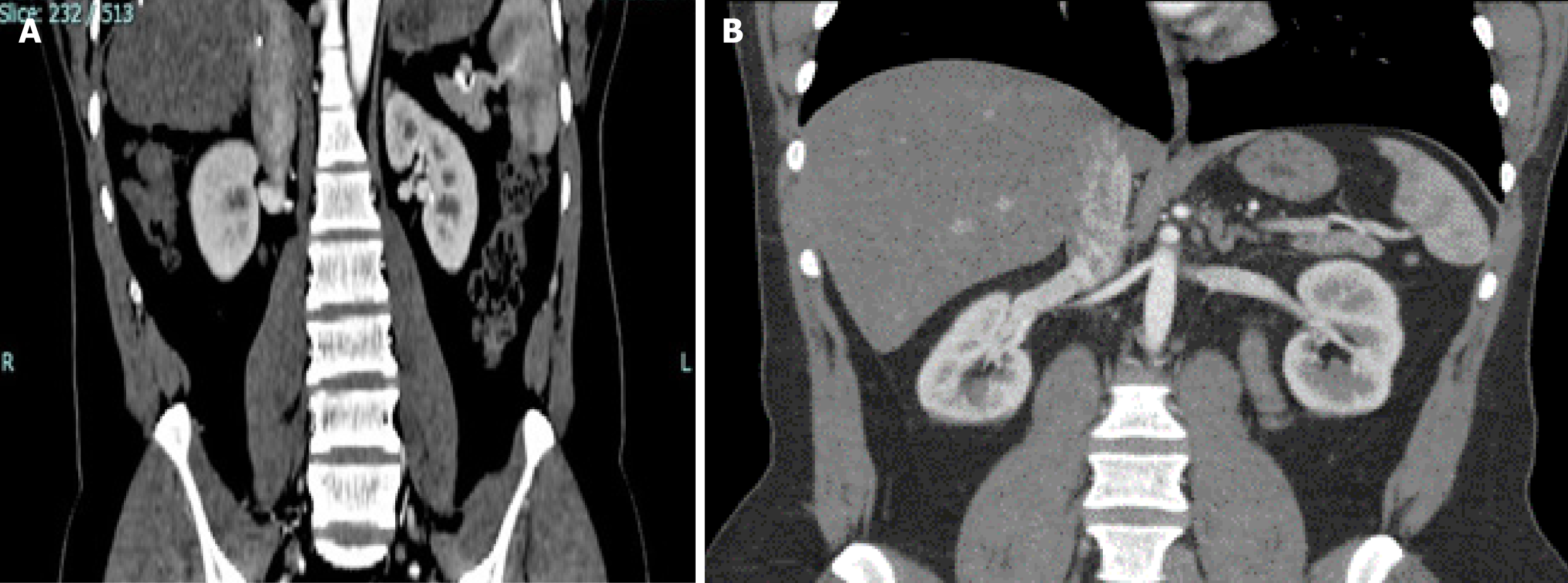
Figure 3 Computed tomography images showing right renal vein entry into the inferior vena cava.
A: Short vein in horizontal entry because perpendicular is the shortest distance; B: Long vein in obtuse angle entry.
- Citation: Khan T, Ahmad N, Iqbal Q, Hassan M, Asnath L, Khan N, Shakeel S. Comparative study of living donor kidney transplants: Right vs left. World J Transplant 2025; 15(1): 97598
- URL: https://www.wjgnet.com/2220-3230/full/v15/i1/97598.htm
- DOI: https://dx.doi.org/10.5500/wjt.v15.i1.97598


