Copyright
©The Author(s) 2024.
World J Transplant. Mar 18, 2024; 14(1): 90277
Published online Mar 18, 2024. doi: 10.5500/wjt.v14.i1.90277
Published online Mar 18, 2024. doi: 10.5500/wjt.v14.i1.90277
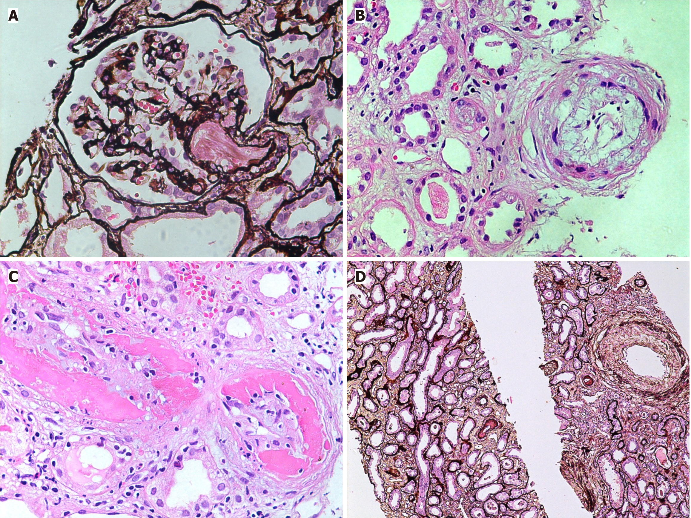
Figure 3 Vascular lesions in thrombotic microangiopathies.
A: Medium-power view showing a glomerulus with an arteriole containing fibrin thrombi in acute phase of thrombotic microangiopathies (TMAs) (H&E, × 200); B: High-power view showing an arteriole with endothelial swelling and complete occlusion of the lumen. An adjacent small artery shows marked mucinous thickening of the intima with narrowing of the lumen (H&E, × 400); C: High-power view showing a small artery with fibrinoid necrosis of the vessel wall and intimal proliferation (H&E, × 400); D: Medium-power view showing fibrointimal thickening of an interlobular size artery in chronic phase of TMA. Mild tubular atrophy is seen in the background (Silver stain, × 200).
- Citation: Mubarak M, Raza A, Rashid R, Sapna F, Shakeel S. Thrombotic microangiopathy after kidney transplantation: Expanding etiologic and pathogenetic spectra. World J Transplant 2024; 14(1): 90277
- URL: https://www.wjgnet.com/2220-3230/full/v14/i1/90277.htm
- DOI: https://dx.doi.org/10.5500/wjt.v14.i1.90277


