Copyright
©The Author(s) 2024.
World J Transplant. Mar 18, 2024; 14(1): 90277
Published online Mar 18, 2024. doi: 10.5500/wjt.v14.i1.90277
Published online Mar 18, 2024. doi: 10.5500/wjt.v14.i1.90277
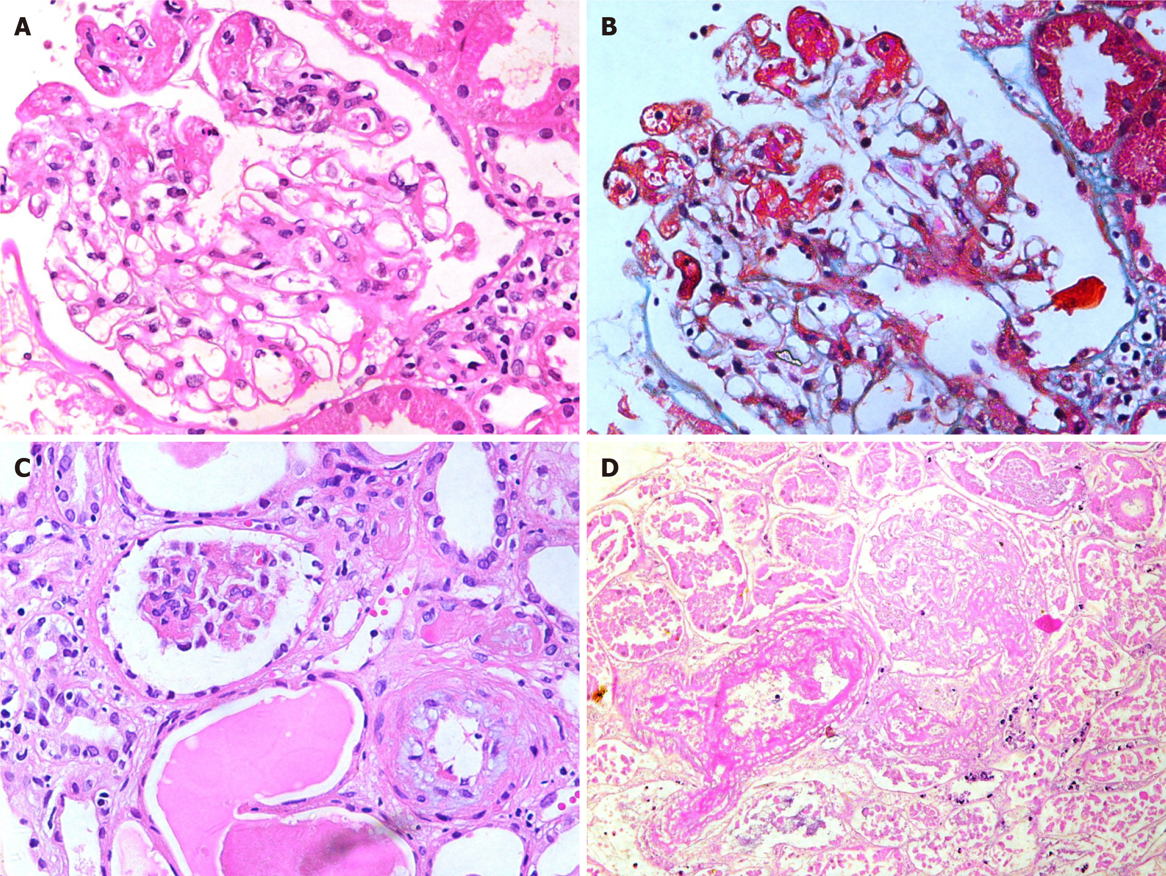
Figure 2 Glomerular lesions in thrombotic microangiopathies.
A: High-power view showing a glomerulus containing fibrin thrombi in dilated capillaries at 9 to 12’O clock position (H&E, × 400); B: The same glomerulus on trichrome staining showing fibrin thrombi staining red with this stain (Masson’s Trichrome, × 400); C: Medium-power view showing one ischemic glomerulus and an arteriole exhibiting mucinous intimal thickening (H&E, × 200); D: Medium-power view showing completely infarcted glomerulus and an adjacent infarcted arteriole containing intraluminal fibrin thrombus. (H&E, × 200).
- Citation: Mubarak M, Raza A, Rashid R, Sapna F, Shakeel S. Thrombotic microangiopathy after kidney transplantation: Expanding etiologic and pathogenetic spectra. World J Transplant 2024; 14(1): 90277
- URL: https://www.wjgnet.com/2220-3230/full/v14/i1/90277.htm
- DOI: https://dx.doi.org/10.5500/wjt.v14.i1.90277


