Copyright
©The Author(s) 2024.
World J Transplant. Mar 18, 2024; 14(1): 88938
Published online Mar 18, 2024. doi: 10.5500/wjt.v14.i1.88938
Published online Mar 18, 2024. doi: 10.5500/wjt.v14.i1.88938
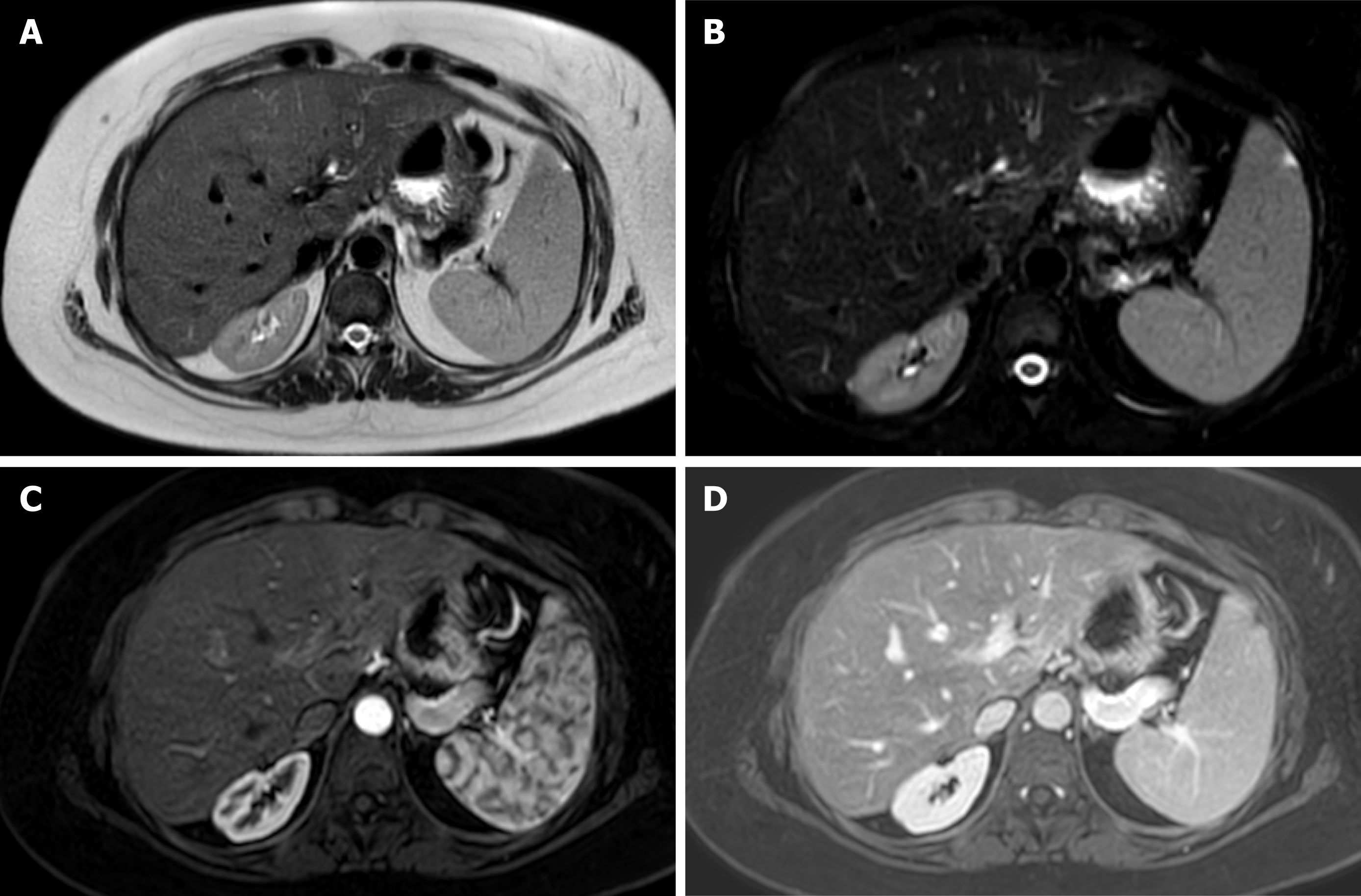
Figure 3 Magnetic resonance imaging of liver graft.
Axial T2-weighted single-shot fast spin-echo image. A: Fat saturation (Fat-sat); B: Contrast enhanced T1-weighted gradient-echo image in late arterial; C: Portal phase; D: Depicts a homogeneous signal intensity at graft parenchyma, with adequate representation of intrahepatic and extrahepatic arterial branches, venous vessels and biliary ducts, in a 32-year-old man on postoperative surveillance after a liver transplantation.
- Citation: Lindner C, Riquelme R, San Martín R, Quezada F, Valenzuela J, Maureira JP, Einersen M. Improving the radiological diagnosis of hepatic artery thrombosis after liver transplantation: Current approaches and future challenges. World J Transplant 2024; 14(1): 88938
- URL: https://www.wjgnet.com/2220-3230/full/v14/i1/88938.htm
- DOI: https://dx.doi.org/10.5500/wjt.v14.i1.88938


