Copyright
©The Author(s) 2023.
World J Transplant. Sep 18, 2023; 13(5): 221-238
Published online Sep 18, 2023. doi: 10.5500/wjt.v13.i5.221
Published online Sep 18, 2023. doi: 10.5500/wjt.v13.i5.221
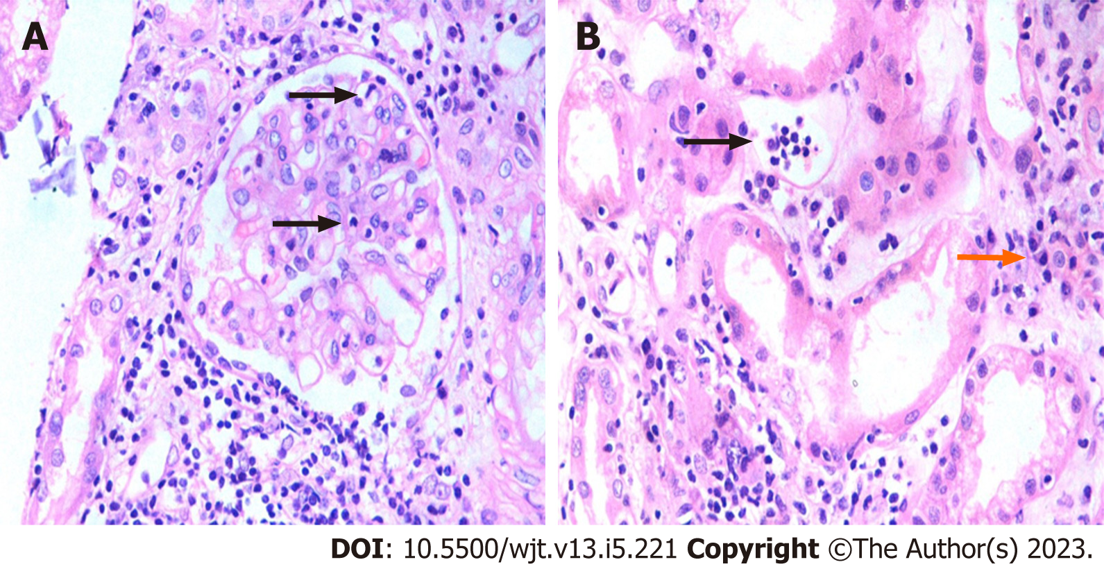
Figure 3 Acute Banff diagnostic lesions of microvascular inflammation.
A: There is segmental glomerulitis (g1) (arrows) along with diffuse interstitial inflammation (i3) in the background [hematoxylin and eosin (H&E), × 400]; B: Peritubular capillaritis (ptc) score ptc 2 (black arrow). ptc is one of the key lesions of antibody-mediated rejection. Patchy interstitial edema, and inflammation with a few plasma cells (orange arrows) are seen in the background (H&E, × 400).
- Citation: Mubarak M, Raza A, Rashid R, Shakeel S. Evolution of human kidney allograft pathology diagnostics through 30 years of the Banff classification process. World J Transplant 2023; 13(5): 221-238
- URL: https://www.wjgnet.com/2220-3230/full/v13/i5/221.htm
- DOI: https://dx.doi.org/10.5500/wjt.v13.i5.221


