Copyright
©The Author(s) 2023.
World J Transplant. Sep 18, 2023; 13(5): 221-238
Published online Sep 18, 2023. doi: 10.5500/wjt.v13.i5.221
Published online Sep 18, 2023. doi: 10.5500/wjt.v13.i5.221
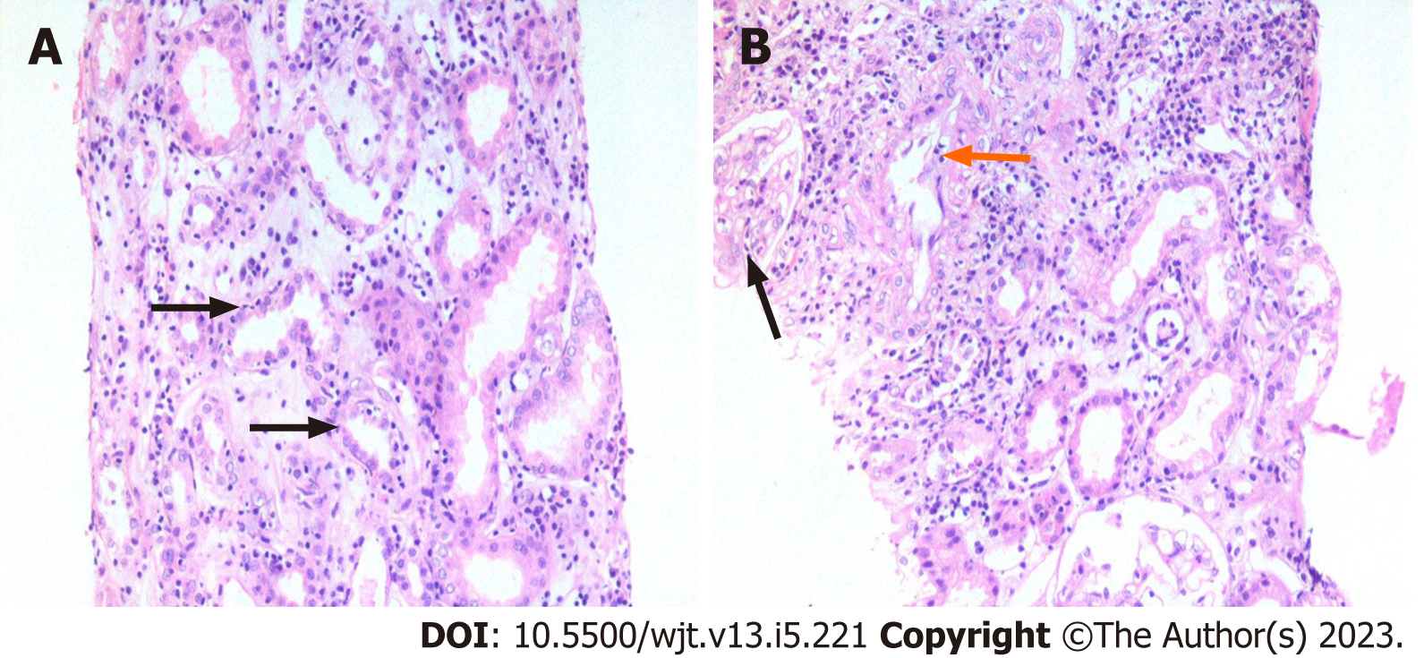
Figure 2 Representative acute Banff diagnostic lesions for rejection and their scores.
A: There is diffuse interstitial inflammation (i3) along with interstitial edema. The later lesion, although important when present, is not formally included in the Banff classification system. Foci of mild tubulitis (t1) are also seen (arrows). These are better visualized on Period acid-Schiff stain [hematoxylin and eosin (H&E), × 200]; B: This field shows glomerulitis (g1) (black arrow) and intimal arteritis (v1) (orange arrow) in addition to i3 and interstitial edema. Such findings raise the suspicion of mixed antibody-mediated and T cell-mediated rejection (H&E, × 200).
- Citation: Mubarak M, Raza A, Rashid R, Shakeel S. Evolution of human kidney allograft pathology diagnostics through 30 years of the Banff classification process. World J Transplant 2023; 13(5): 221-238
- URL: https://www.wjgnet.com/2220-3230/full/v13/i5/221.htm
- DOI: https://dx.doi.org/10.5500/wjt.v13.i5.221


