Copyright
©The Author(s) 2021.
World J Transplant. Jun 18, 2021; 11(6): 231-243
Published online Jun 18, 2021. doi: 10.5500/wjt.v11.i6.231
Published online Jun 18, 2021. doi: 10.5500/wjt.v11.i6.231
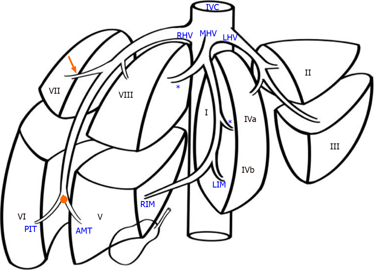
Figure 1 Variations of the right hepatic vein.
In the normal pattern, the left hepatic vein (LHV) runs within the inter-sectional plane between segments II and III, continuing to enter the inferior vena cava (IVC). The middle hepatic vein (MHV) is formed at the union of the left inferior middle (LIM) branch vein and the right inferior middle (RIM) vein branch. It travels cephalad in the mid-plane of the liver to enter the IVC, and receives tributaries from both halves of liver, known as left and right superior middle vein branches (asterisk). The right hepatic vein (RHV) forms at the hepatic venous confluence (orange dot) where two large tributaries, meet: the anteromedial tributary (AMT) that drains segments V and the posterioinferior tributary (PIT) that draining segment VI, meet. The two tributaries join to form the superior RHV that then courses up toward the IVC. A tributary draining segment VII (orange arrow) consistently joins the superior RHV from its posterolateral side.
- Citation: Cawich SO, Naraynsingh V, Pearce NW, Deshpande RR, Rampersad R, Gardner MT, Mohammed F, Dindial R, Barrow TA. Surgical relevance of anatomic variations of the right hepatic vein. World J Transplant 2021; 11(6): 231-243
- URL: https://www.wjgnet.com/2220-3230/full/v11/i6/231.htm
- DOI: https://dx.doi.org/10.5500/wjt.v11.i6.231


