Copyright
©The Author(s) 2021.
World J Transplant. Jun 18, 2021; 11(6): 161-179
Published online Jun 18, 2021. doi: 10.5500/wjt.v11.i6.161
Published online Jun 18, 2021. doi: 10.5500/wjt.v11.i6.161
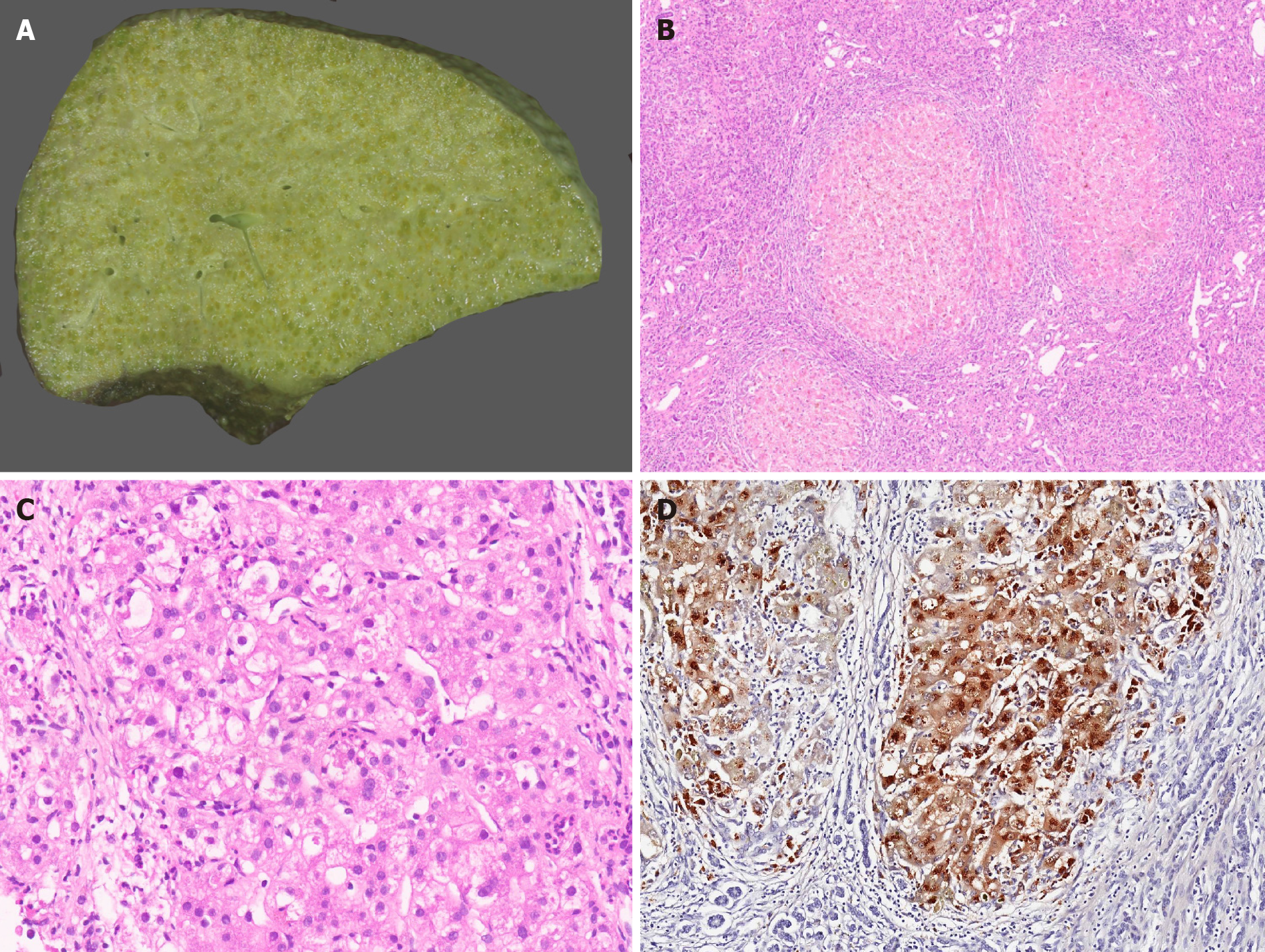
Figure 2 Wilson disease.
A: Explant hepatectomy specimen in a case of Wilson disease (WD) with marked cholestasis; B: Micronodular cirrhosis in a case of WD [hematoxylin and eosin (HE staining)]; C: WD with hepatocellular ballooning, Mallory Denk bodies, fatty change and neutrophilic satellitosis (HE staining); and D: WD with marked copper deposition in hepatocytes and macrophages (Rhodanine staining).
- Citation: Menon J, Vij M, Sachan D, Rammohan A, Shanmugam N, Kaliamoorthy I, Rela M. Pediatric metabolic liver diseases: Evolving role of liver transplantation. World J Transplant 2021; 11(6): 161-179
- URL: https://www.wjgnet.com/2220-3230/full/v11/i6/161.htm
- DOI: https://dx.doi.org/10.5500/wjt.v11.i6.161


