Copyright
©The Author(s) 2020.
World J Transplant. May 29, 2020; 10(5): 129-137
Published online May 29, 2020. doi: 10.5500/wjt.v10.i5.129
Published online May 29, 2020. doi: 10.5500/wjt.v10.i5.129
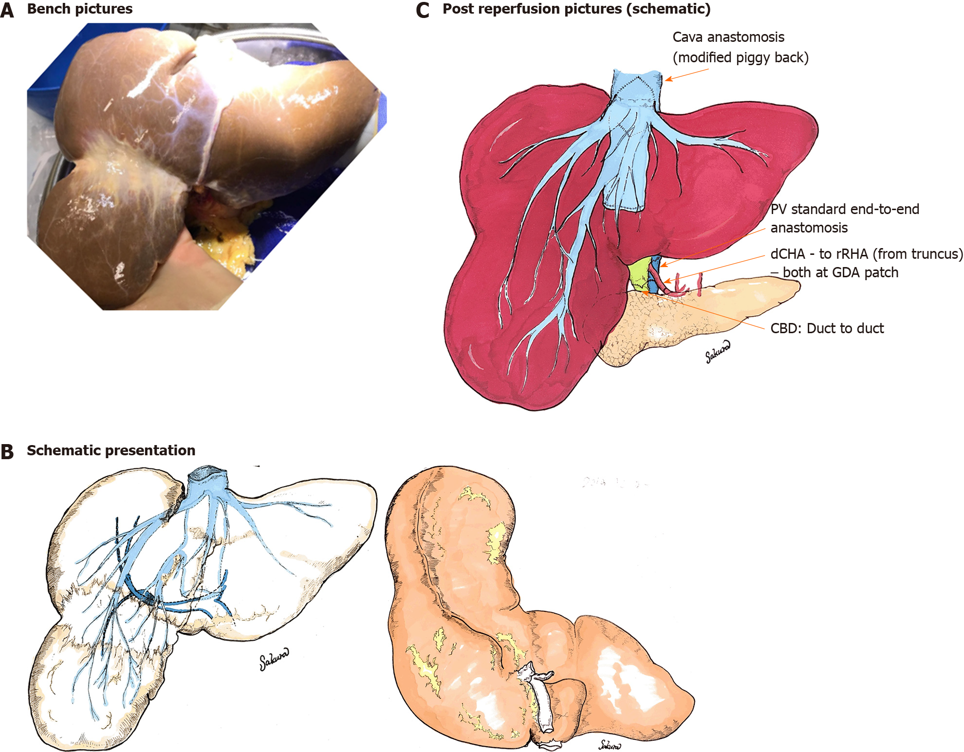
Figure 3 Schematic overview of donor liver with Riedel’s lobe (Case 2): Bench picture.
A: Schematic presentation of the liver; B: With large segmental separation of S6/7 and 5/8, a special variation of a Riedel’s lobe and the implantation technique; C: The implantation was performed with a side to side piggy back technique in a routine way with longitudinal incision of the donor and recipient inferior vena cava. This was in contrast to the first case. PV: Portal vein; CHA: Common hepatic artery; RHA: Right hepatic artery; GDA: Gastroduodenal artery; CBD: Common bile duct.
- Citation: Sakuraoka Y, Seth R, Boteon AP, Perrin M, Isaac J, Subash G, Muiesan P, Schlegel A. Large Riedel’s lobe and atrophic left liver in a donor - Accept for transplant or call off? World J Transplant 2020; 10(5): 129-137
- URL: https://www.wjgnet.com/2220-3230/full/v10/i5/129.htm
- DOI: https://dx.doi.org/10.5500/wjt.v10.i5.129


