Copyright
©2014 Baishideng Publishing Group Inc.
World J Med Genet. Nov 27, 2014; 4(4): 77-93
Published online Nov 27, 2014. doi: 10.5496/wjmg.v4.i4.77
Published online Nov 27, 2014. doi: 10.5496/wjmg.v4.i4.77
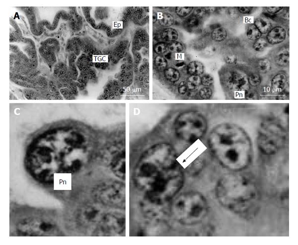
Figure 7 Silver fox placenta.
A and B: Trabeculae of trophoblast and folds of uterine glandular epithelium (Ep) mutually contact each other, trophoblast giant cells (TGC) are scattered in the fetal part of placenta between accumulations of proliferative cells; B: Mitotic (M), binucleate (Bc) and cells with polytene nuclei (Pn); C: Nucleus with non-classic polytene chromosomes; D: A polyploid nucleus in the beginning of fragmentation (arrow). Meyer hematoxylin staining.
- Citation: Zybina TG, Zybina EV. Genome variation in the trophoblast cell lifespan: Diploidy, polyteny, depolytenization, genome segregation. World J Med Genet 2014; 4(4): 77-93
- URL: https://www.wjgnet.com/2220-3184/full/v4/i4/77.htm
- DOI: https://dx.doi.org/10.5496/wjmg.v4.i4.77


