Copyright
©2012 Baishideng.
World J Clin Infect Dis. Feb 25, 2012; 2(1): 1-12
Published online Feb 25, 2012. doi: 10.5495/wjcid.v2.i1.1
Published online Feb 25, 2012. doi: 10.5495/wjcid.v2.i1.1
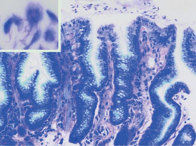
Figure 1 Histological examination of a gastric lesion from an infected patient showing multiple Giardia intestinalis trophozoites (10-20 μm × 5-15 μm) adhering to the surface epithelium (magnification x 200, Giemsa stain).
Magnification × 1000 (inset) reveals the typical tear-drop shape with twin nuclei of this parasite. Image reproduced with kind permission from Georg Thieme Verlag KG, Stuttgart, Germany.
- Citation: Carmena D, Cardona GA, Sánchez-Serrano LP. Current situation of Giardia infection in Spain: Implications for public health. World J Clin Infect Dis 2012; 2(1): 1-12
- URL: https://www.wjgnet.com/2220-3176/full/v2/i1/1.htm
- DOI: https://dx.doi.org/10.5495/wjcid.v2.i1.1


