Copyright
©The Author(s) 2015.
World J Hypertens. May 23, 2015; 5(2): 28-40
Published online May 23, 2015. doi: 10.5494/wjh.v5.i2.28
Published online May 23, 2015. doi: 10.5494/wjh.v5.i2.28
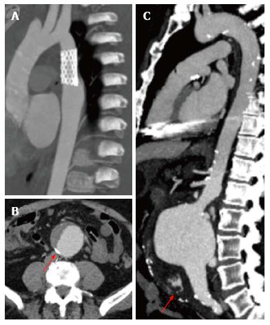
Figure 8 Aortic computed tomography.
A: Patient with vascular endoprothesis, without vascular leaks; B: Dissected abdominal aortic aneurysm, arrow points to calcified atherosclerotic plaques; C: Ruptured infrarenal aortic aneurysm, seen as hyperdense material in the abdominal cavity (arrow).
- Citation: Alexanderson-Rosas E, Berríos-Bárcenas E, Meave A, de la Fuente-Mancera JC, Oropeza-Aguilar M, Barrero-Mier A, Monroy-González AG, Cruz-Mendoza R, Guinto-Nishimura GY. Novel contributions of multimodality imaging in hypertension: A narrative review. World J Hypertens 2015; 5(2): 28-40
- URL: https://www.wjgnet.com/2220-3168/full/v5/i2/28.htm
- DOI: https://dx.doi.org/10.5494/wjh.v5.i2.28


