Copyright
©The Author(s) 2015.
World J Hypertens. May 23, 2015; 5(2): 28-40
Published online May 23, 2015. doi: 10.5494/wjh.v5.i2.28
Published online May 23, 2015. doi: 10.5494/wjh.v5.i2.28
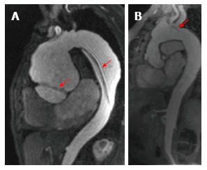
Figure 4 Evaluation of vascular anatomy using cardiovascular magnetic resonance.
A: Patient with Marfan’s syndrome that presents a Stanford A type dissected aortic aneurysm; the arrows point to the two sites of dissection; B shows post-surgical changes after a Bentall and Bono procedure; the arrow points to a dissection flap in the aortic arch.
- Citation: Alexanderson-Rosas E, Berríos-Bárcenas E, Meave A, de la Fuente-Mancera JC, Oropeza-Aguilar M, Barrero-Mier A, Monroy-González AG, Cruz-Mendoza R, Guinto-Nishimura GY. Novel contributions of multimodality imaging in hypertension: A narrative review. World J Hypertens 2015; 5(2): 28-40
- URL: https://www.wjgnet.com/2220-3168/full/v5/i2/28.htm
- DOI: https://dx.doi.org/10.5494/wjh.v5.i2.28


