Copyright
©The Author(s) 2015.
World J Hypertens. May 23, 2015; 5(2): 28-40
Published online May 23, 2015. doi: 10.5494/wjh.v5.i2.28
Published online May 23, 2015. doi: 10.5494/wjh.v5.i2.28
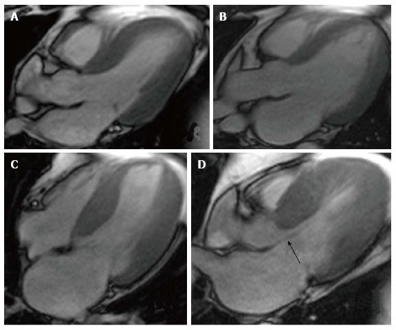
Figure 1 Cardiovascular magnetic resonance showing Steady-state Free Precision Sequences.
A: Patient with hypertension presenting slight ventricular hypertrophy (septal wall of 13 mm); B: Patient with hypertensive cardiomyopathy in dilated phase with a LVDD = 70 mm, LVEF = 40%, left atrium = 65 mm; C: 51-year-old male patient with asymmetrical septal hypertrophy, with a maximum thickness of 27 mm; D: Same patient as in C, the arrow shows the anterior systolic movement of the mitral valve, generating outflow tract obstruction.
- Citation: Alexanderson-Rosas E, Berríos-Bárcenas E, Meave A, de la Fuente-Mancera JC, Oropeza-Aguilar M, Barrero-Mier A, Monroy-González AG, Cruz-Mendoza R, Guinto-Nishimura GY. Novel contributions of multimodality imaging in hypertension: A narrative review. World J Hypertens 2015; 5(2): 28-40
- URL: https://www.wjgnet.com/2220-3168/full/v5/i2/28.htm
- DOI: https://dx.doi.org/10.5494/wjh.v5.i2.28


