Copyright
©The Author(s) 2025.
World J Exp Med. Jun 20, 2025; 15(2): 100166
Published online Jun 20, 2025. doi: 10.5493/wjem.v15.i2.100166
Published online Jun 20, 2025. doi: 10.5493/wjem.v15.i2.100166
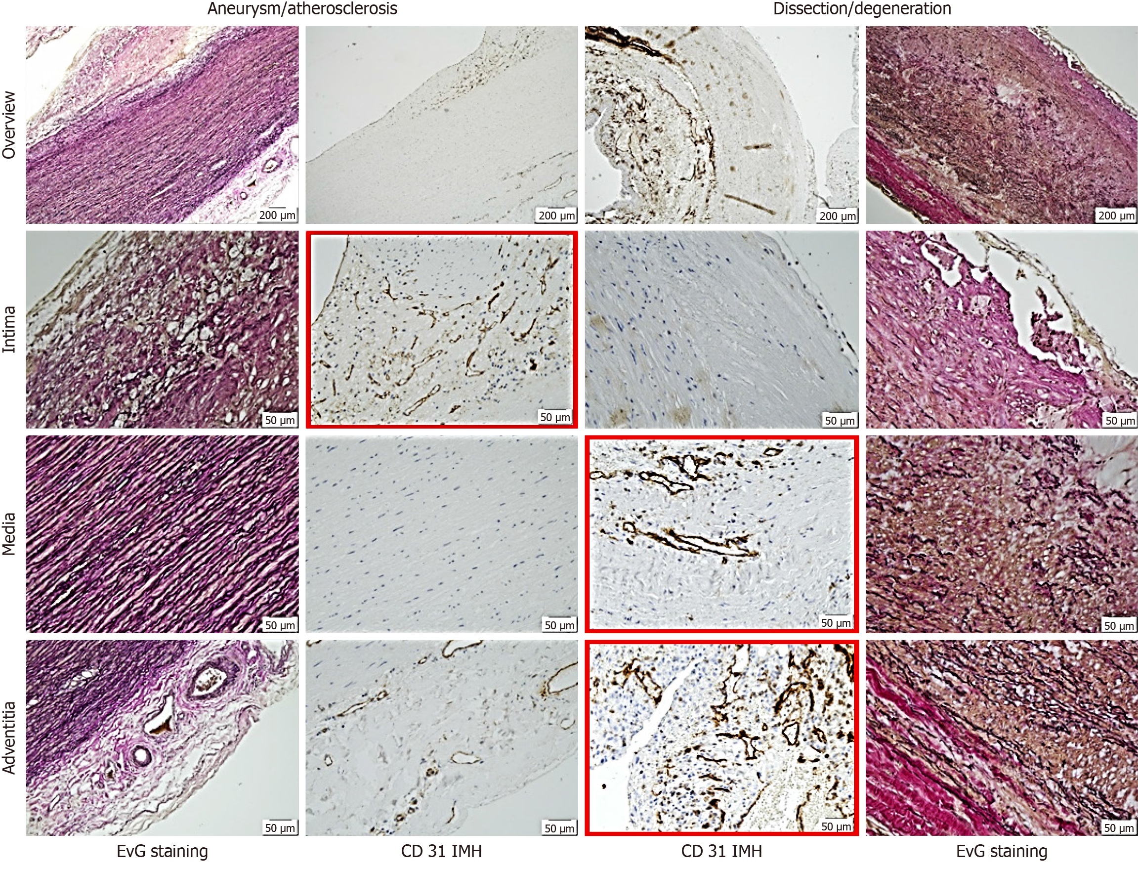
Figure 2 Representative histological and immunohistological findings in aneurysm and dissection.
Note the differing staining patterns of vessels: In the intima for aneurysmal changes and in the media and adventitia for dissection (red frames). The distinct pattern of small vessels reflects vascularization or neovascularization, suggesting the primarily degenerative nature of dissection pathogenesis. Staining methods included hematoxylin (S3301, Dako), Elastica van Gieson, and immunohistochemistry using a primary antibody (monoclonal Mouse Anti-human CD31, Endothelial Cell Clone JC70A, Dako). EvG: Elastica van Gieson.
- Citation: Irqsusi M, Rodepeter FR, Günther M, Kirschbaum A, Vogt S. Matrix metalloproteinases and their tissue inhibitors as indicators of aortic aneurysm and dissection development in extracellular matrix remodeling. World J Exp Med 2025; 15(2): 100166
- URL: https://www.wjgnet.com/2220-315x/full/v15/i2/100166.htm
- DOI: https://dx.doi.org/10.5493/wjem.v15.i2.100166


