Copyright
©The Author(s) 2021.
World J Exp Med. Dec 30, 2021; 11(6): 79-92
Published online Dec 30, 2021. doi: 10.5493/wjem.v11.i6.79
Published online Dec 30, 2021. doi: 10.5493/wjem.v11.i6.79
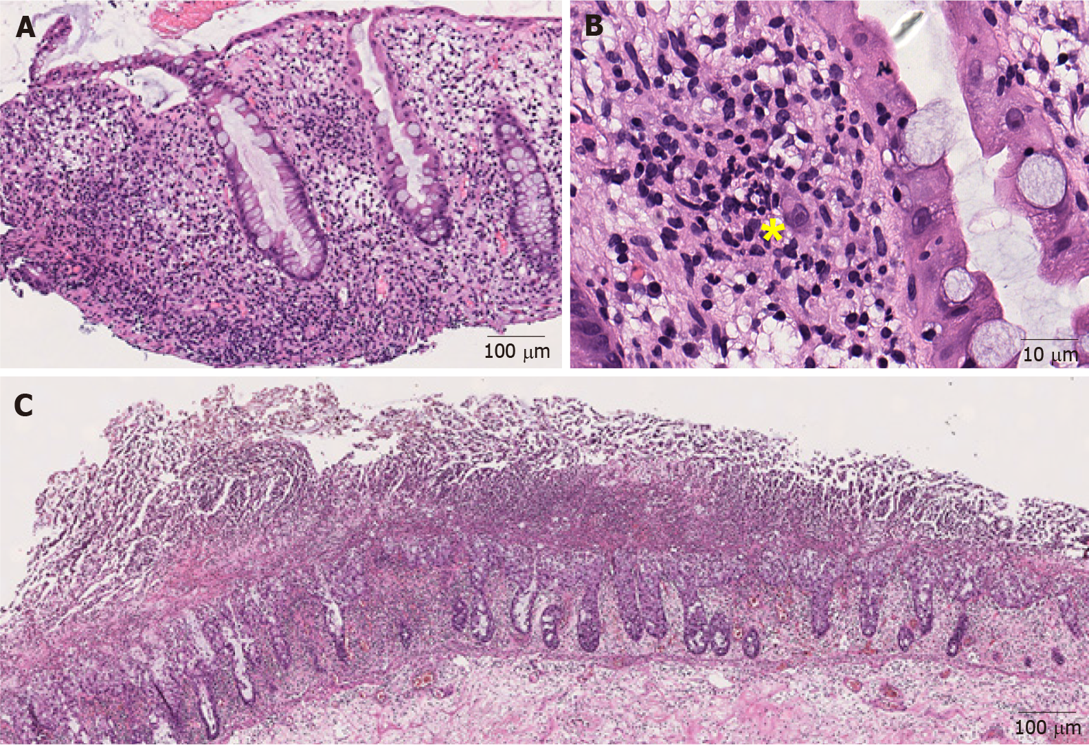
Figure 2 Representative images of infectious colitis (Hematoxylin and eosin).
A: Low magnification view of cytomegaloviral (CMV) colitis. Notice the lymphocytes and neutrophils in the lamina propria (100 ×); B: Note an owl-eye inclusion characterized by enlarged nucleus with oval, eosinophilic intranuclear inclusion surrounded by clear halo, consistent with CMV inclusion (400 ×); C: Clostridium difficile colitis. Pseudomembranes composed of fibrin, neutrophils and necrotic epithelial cells are on the surface of the mucosal glands (40 ×). Yellow sign notes a viral inclusiona.
- Citation: Li H, Fu ZY, Arslan ME, Cho D, Lee H. Differential diagnosis and management of immune checkpoint inhibitor-induced colitis: A comprehensive review. World J Exp Med 2021; 11(6): 79-92
- URL: https://www.wjgnet.com/2220-315x/full/v11/i6/79.htm
- DOI: https://dx.doi.org/10.5493/wjem.v11.i6.79


