Copyright
©The Author(s) 2019.
World J Exp Med. Dec 19, 2019; 9(2): 14-31
Published online Dec 19, 2019. doi: 10.5493/wjem.v9.i2.14
Published online Dec 19, 2019. doi: 10.5493/wjem.v9.i2.14
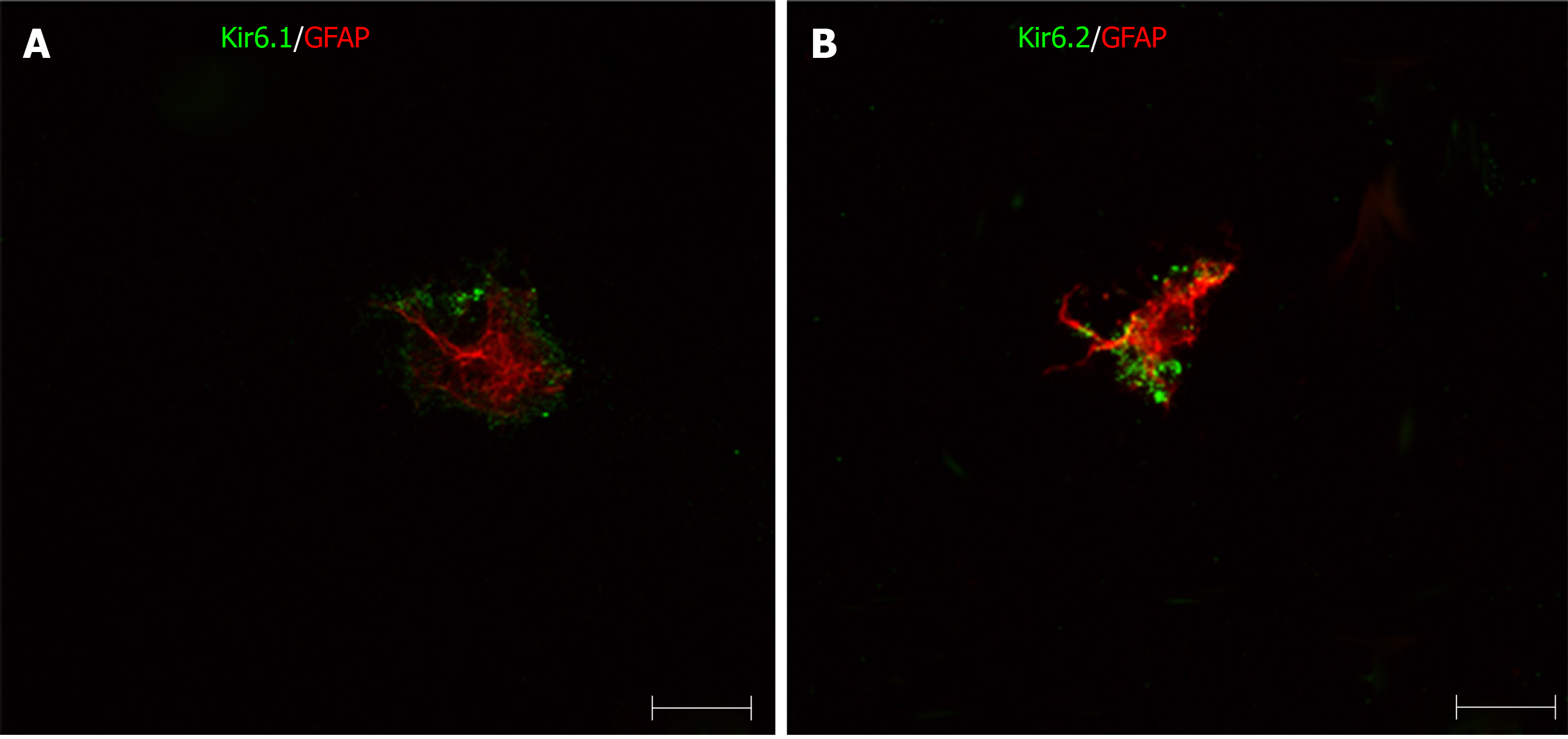
Figure 13 Immunofluorescence double staining for pore-forming subunits in primary cultured hepatic stellate cells.
The primary hepatic stellate cells were marked by GFAP (red), they showed immunoreactivity to Kir6.1 and Kir6.2 (green). Bars: 50 µm.
- Citation: Zhou M, Yoshikawa K, Akashi H, Miura M, Suzuki R, Li TS, Abe H, Bando Y. Localization of ATP-sensitive K+ channel subunits in rat liver. World J Exp Med 2019; 9(2): 14-31
- URL: https://www.wjgnet.com/2220-315X/full/v9/i2/14.htm
- DOI: https://dx.doi.org/10.5493/wjem.v9.i2.14


