Copyright
©The Author(s) 2017.
World J Exp Med. Aug 20, 2017; 7(3): 84-96
Published online Aug 20, 2017. doi: 10.5493/wjem.v7.i3.84
Published online Aug 20, 2017. doi: 10.5493/wjem.v7.i3.84
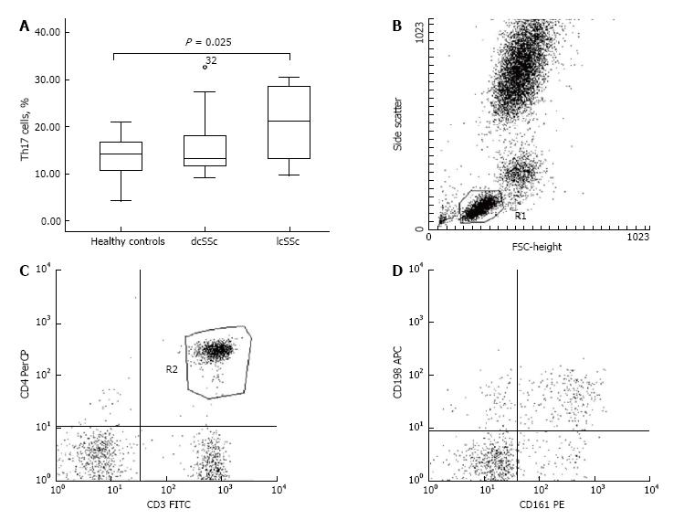
Figure 2 Increased percentage of Th17 cells within limited cutaneous systemic sclerosis subset vs healthy controls.
A: Percentage of Th17 cells for lcSSc (n = 11), dcSSc (n = 13), and healthy controls (n = 16) is presented. Increased percentage of Th17 cells within lcSSc patients as opposed to controls, respectively, 20.46% ± 2.41% vs 13.73% ± 1.21%, P = 0.025. Boxplots are expressed as means ± SD; B-D: Panel B depicts the flow cytometric analysis of Th17 cells. A representative patient with lcSSc phenotype is shown. The lymphocytes were gated according to their physical characteristics (FSC and SSC) in R1; afterwards T helper cells (CD3+CD4+) were gated in R2. T helpers, which were detected double positive for CD161 and CD196 surface expression (R3, upper right quadrant) were defined as Th17 cells. lcSSc: Limited cutaneous systemic sclerosis.
- Citation: Krasimirova E, Velikova T, Ivanova-Todorova E, Tumangelova-Yuzeir K, Kalinova D, Boyadzhieva V, Stoilov N, Yoneva T, Rashkov R, Kyurkchiev D. Treg/Th17 cell balance and phytohaemagglutinin activation of T lymphocytes in peripheral blood of systemic sclerosis patients. World J Exp Med 2017; 7(3): 84-96
- URL: https://www.wjgnet.com/2220-315X/full/v7/i3/84.htm
- DOI: https://dx.doi.org/10.5493/wjem.v7.i3.84


