Copyright
©The Author(s) 2016.
World J Exp Med. Nov 20, 2016; 6(4): 72-79
Published online Nov 20, 2016. doi: 10.5493/wjem.v6.i4.72
Published online Nov 20, 2016. doi: 10.5493/wjem.v6.i4.72
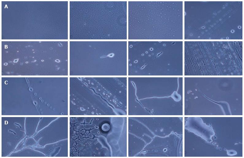
Figure 2 Phase contrast images of neuronal cells and tissues at different days of culture.
A: Day 0: Small stem cells; B: Day 5: Differentiating neurons; C: Day 16-18: Small protrusions, neuritis structure and end-joining of neurons; D: Day 30: Complete neurons and neuronal tissue.
- Citation: Shroff G. Morphogenesis of human embryonic stem cells into mature neurons under in vitro culture conditions. World J Exp Med 2016; 6(4): 72-79
- URL: https://www.wjgnet.com/2220-315X/full/v6/i4/72.htm
- DOI: https://dx.doi.org/10.5493/wjem.v6.i4.72


