Copyright
©The Author(s) 2015.
World J Exp Med. Aug 20, 2015; 5(3): 164-181
Published online Aug 20, 2015. doi: 10.5493/wjem.v5.i3.164
Published online Aug 20, 2015. doi: 10.5493/wjem.v5.i3.164
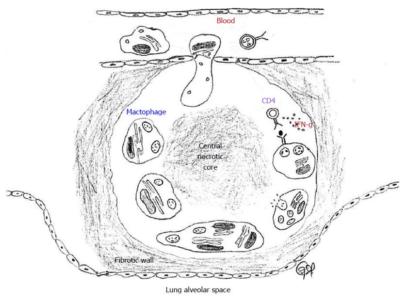
Figure 3 Tuberculosis granuloma.
A granuloma sequesters MTB infected macrophages and is surrounded by immune cells, predominantly CD4+ helper T lymphocytes. Some infected macrophages fuse to form foamy giant cells. The infected macrophages and giant cells present antigens to T cells and activate them to produce a variety of cytokines and chemokynes, and also kills the infected macrophage and the MTB. The chemokines also serve to recruit additional T cells to the granuloma from the circulating blood. IFN-γ activates the macrophages to kill the intracellular MTB by generating reactive oxygen species and reactive nitrogen species intracellularly. The center of the granuloma is filled with cell debris and both live and dead MTB spilled from dead macrophages (caseation), all of which form a central hypoxic necrotic core. A sheath of collagen fibers produced from lung fibroblasts surround the granuloma. MTB: Mycobacterium tuberculosis.
- Citation: George R, Cavalcante R, Jr CC, Marques E, Waugh JB, Unlap MT. Use of siRNA molecular beacons to detect and attenuate mycobacterial infection in macrophages. World J Exp Med 2015; 5(3): 164-181
- URL: https://www.wjgnet.com/2220-315X/full/v5/i3/164.htm
- DOI: https://dx.doi.org/10.5493/wjem.v5.i3.164


